. 2014 Oct 28;30(42):12610-8.
doi: 10.1021/la502610c. Epub 2014 Oct 13.

Recombinant protein-stabilized monodisperse microbubbles with tunable size using a valve-based microfluidic device

Affiliations

Recombinant protein-stabilized monodisperse microbubbles with tunable size using a valve-based microfluidic device

Francesco E Angilè et al. Langmuir. .

Abstract

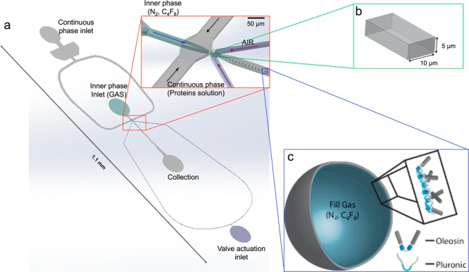
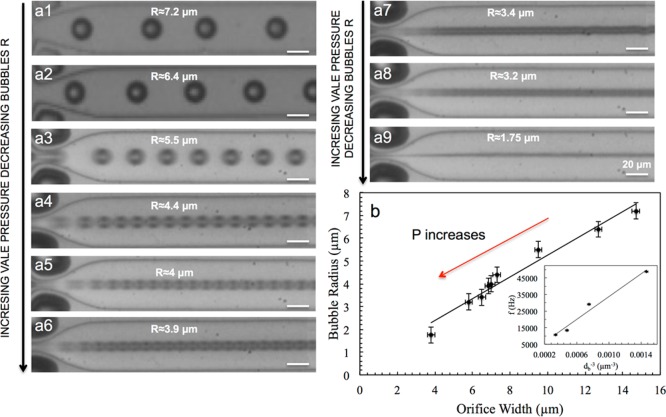
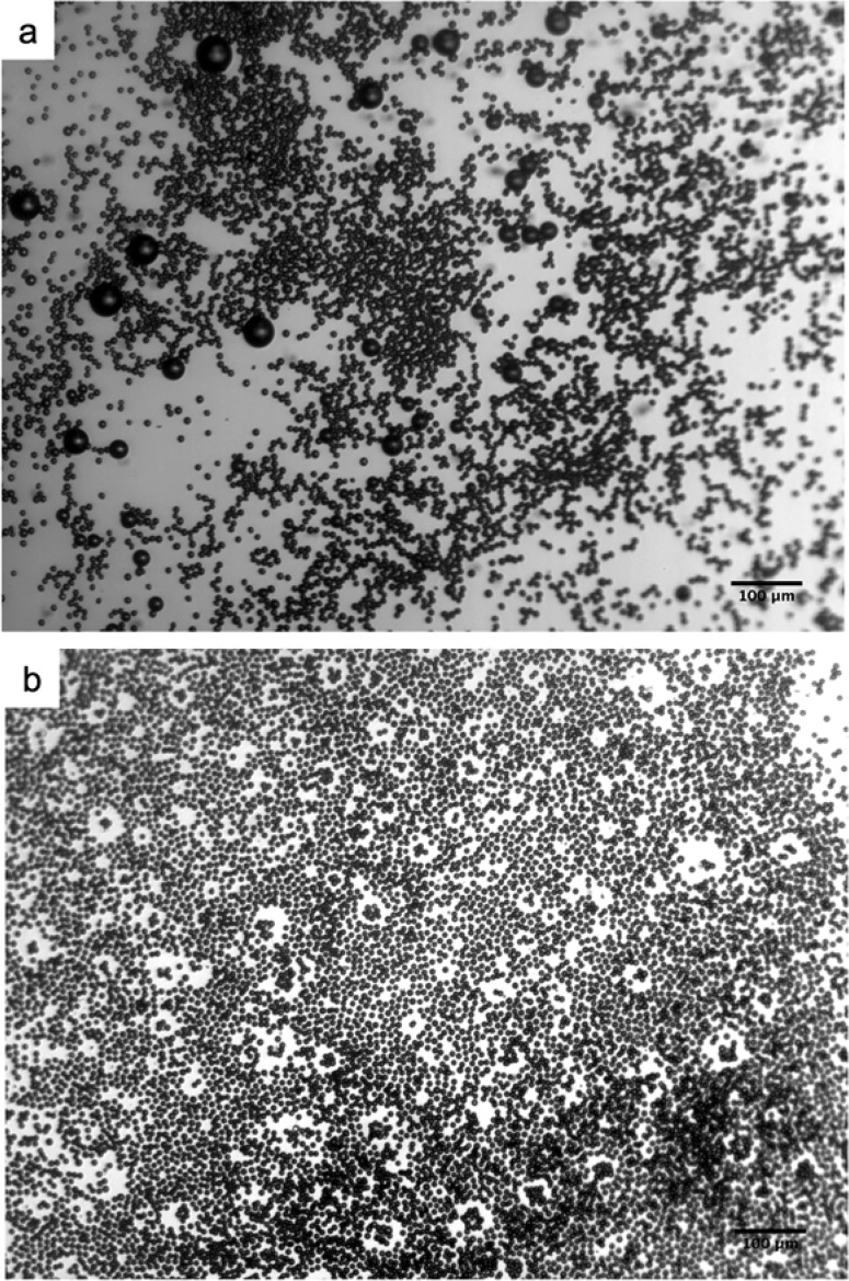
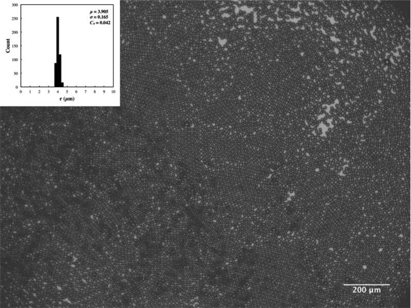
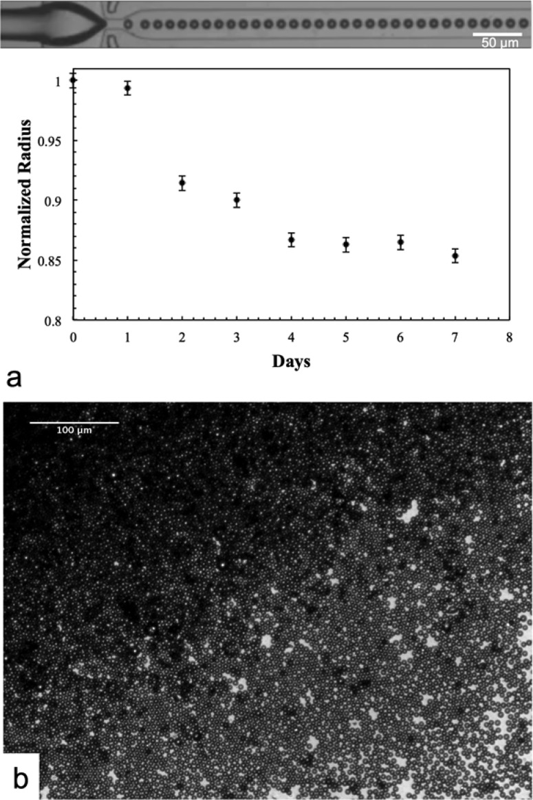
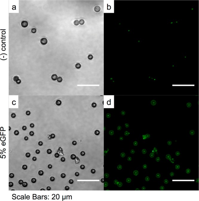
Microbubbles are used as contrast enhancing agents in ultrasound sonography and more recently have shown great potential as theranostic agents that enable both diagnostics and therapy. Conventional production methods lead to highly polydisperse microbubbles, which compromise the effectiveness of ultrasound imaging and therapy. Stabilizing microbubbles with surfactant molecules that can impart functionality and properties that are desirable for specific applications would enhance the utility of microbubbles. Here we generate monodisperse microbubbles with a large potential for functionalization by combining a microfluidic method and recombinant protein technology. Our microfluidic device uses an air-actuated membrane valve that enables production of monodisperse microbubbles with narrow size distribution. The size of microbubbles can be precisely tuned by dynamically changing the dimension of the channel using the valve. The microbubbles are stabilized by an amphiphilic protein, oleosin, which provides versatility in controlling the functionalization of microbubbles through recombinant biotechnology. We show that it is critical to control the composition of the stabilizing agents to enable formation of highly stable and monodisperse microbubbles that are echogenic under ultrasound insonation. Our protein-shelled microbubbles based on the combination of microfluidic generation and recombinant protein technology provide a promising platform for ultrasound-related applications.


Figures

Figure 1
Figure 1
(a) Schematic illustration of a PDMS microfluidic device used to generate monodisperse microbubbles of different sizes. (b) Cross-sectional geometry of the nozzle (see Figure S1 for junction dimensions). (c) Schematic of a microbubble stabilized with a mixture of oleosin and (PEO)n-(PPO)m-(PEO)n triblock copolymer.
Figure 2
Figure 2
(a1–a9) Series of micrographs of the microfluidic device during the generation of microbubbles using a solution containing SDS at a concentration of 20 mg mL–1 in the aqueous phase. By changing the size of the nozzle, which is controlled by an air-actuated valve placed at the orifice, it is possible to generate uniform microbubbles of different sizes. (b) Effect of orifice width on the size of microbubbles. The inset shows the microbubbles generation frequency (f) vs volume of microbubbles (db–3). The linear relationship between the two quantities indicates that the gas flow rates remains more or less constant under varying nozzle size.
Figure 3
Figure 3
(a) SDS-PAGE gel showing >95% purity for 42-30G-63. (b) MADLI-TOF spectra confirming the molecular weight for 42-30G-63 (expected: 15 027; measured: 15 025). (c) Far-ultraviolet circular dichroism (UV CD) spectra of 42-30G-63 and wild-type oleosin. The former indicates a random coil structure.
Figure 4
Figure 4
FTIR absorbance spectra of the components utilized to produce the bubbles and microbubbles. The spectra of pure oleosin and microbubbles are amplified by factors of 2.5 and 5, respectively, to clearly show the features.
Figure 5
Figure 5
Micrographs of microbubbles produced using a solution containing 1 mg mL1 oleosin and 10 mg mL–1 (PEO)78-(PPO)30-(PEO)78. (a) A small number of large bubbles are present upon collection via plastic tubing. (b) Big bubbles disappear 24 h after collection, leaving monodisperse microbubbles.
Figure 6
Figure 6
Micrograph of monodisperse microbubbles produced using a solution containing 1 mg mL1 oleosin and 10 mg mL–1 (PEO)78-(PPO)30-(PEO)78 and collected into a well in the PDMS device without the use of plastic tubing. The inset shows the microbubble size distribution for ∼500 microbubbles. μ, σ, and Cv in the inset represent the average (in μm), standard deviation (in μm), and coefficient of variation, respectively.
Figure 7
Figure 7
Micrographs showing microbubbles stability over time for microbubbles produced using a solution containing 1 mg mL1 oleosin and 10 mg mL–1 (PEO)78-(PPO)30-(PEO)78. (a) Size of microbubbles over 7 days. (b) Microscope images of 24 days after collection.
Figure 8
Figure 8
Confocal fluorescent microscopy images of bubbles produced with (a, b) oleosin and (c, d) with a blend containing the eGFP mutant. In both cases a solution containing 1 mg mL1 oleosin and 10 mg mL–1 (PEO)78-(PPO)30-(PEO)78 is used to produce microbubbles. Microbubbles are stored for 24 h before confocal microscopy is performed. These images are taken by focusing at the equatorial planes of the bubbles.
Figure 9
Figure 9
Ultrasound sonography images of C4F8 microbubbles generated with a solution containing 1 mg mL1 oleosin and 10 mg mL–1 (PEO)78-(PPO)30-(PEO)78. Ultrasound images of microbubbles (a, b) 1–2 h after generation and (c, d) 30 min and (e, f) 7 days after initial imaging. Ultrasound images of control samples are reported in panels g and h. The microbubbles have a radius of about 4 μm.

References

    1. Kiessling F.; Fokong S.; Koczera P.; Lederle W.; Lammers T. Ultrasound microbubbles for molecular diagnosis, therapy, and theranostics. J. Nucl. Med. 2012, 53, 345–348. - PubMed
    1. de Jong N.; Cate F. T.; Lancée C.; Roelandt J.; Bom N. Principles and recent developments in ultrasound contrast agents. Ultrasonics 1991, 29, 324–330. - PubMed
    1. Del Vecchio S.; Zannetti A.; Fonti R. Nuclear Imaging in cancer theranostic. Q. J. Nucl. Med. Mol. Imaging 2007, 51, 152–163. - PubMed
    1. Segers T.; Versluis M. Acoustic bubble sorting for ultrasound contrast agent enrichment. Lab Chip 2014, 14, 1705–1714. - PubMed
    1. Talu E.; Hettiarachchi K.; Zhao S.; Powell R. L.; Lee A. P.; Longo L.; Dayton P. A. Tailoring the size distribution of ultrasound contrast agents: possible method for improving sensitivity in molecular imaging. Mol. Imaging 2007, 6, 384–392. - PMC - PubMed

Publication types

MeSH terms

LinkOut - more resources