Review
. 2015 Jan:11:3-16.
doi: 10.1016/j.actbio.2014.09.038. Epub 2014 Oct 5.

Evolving insights in cell-matrix interactions: elucidating how non-soluble properties of the extracellular niche direct stem cell fate

Affiliations
Review

Evolving insights in cell-matrix interactions: elucidating how non-soluble properties of the extracellular niche direct stem cell fate

Nick J Walters et al. Acta Biomater. 2015 Jan.

Abstract

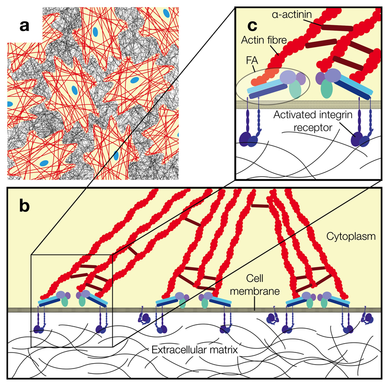
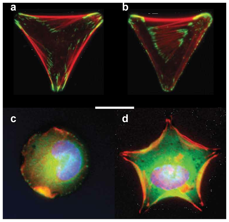
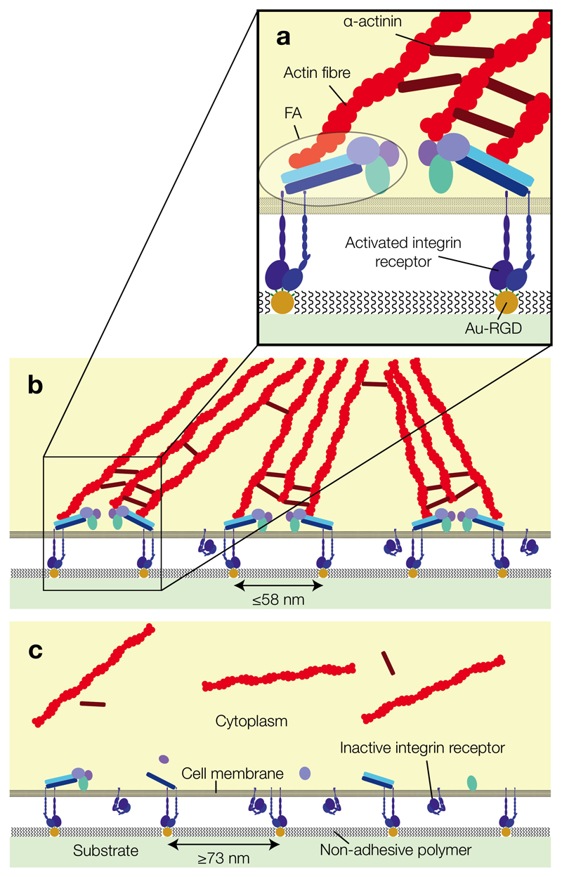
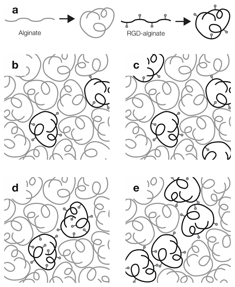
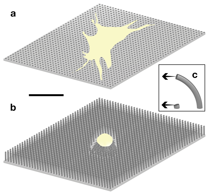
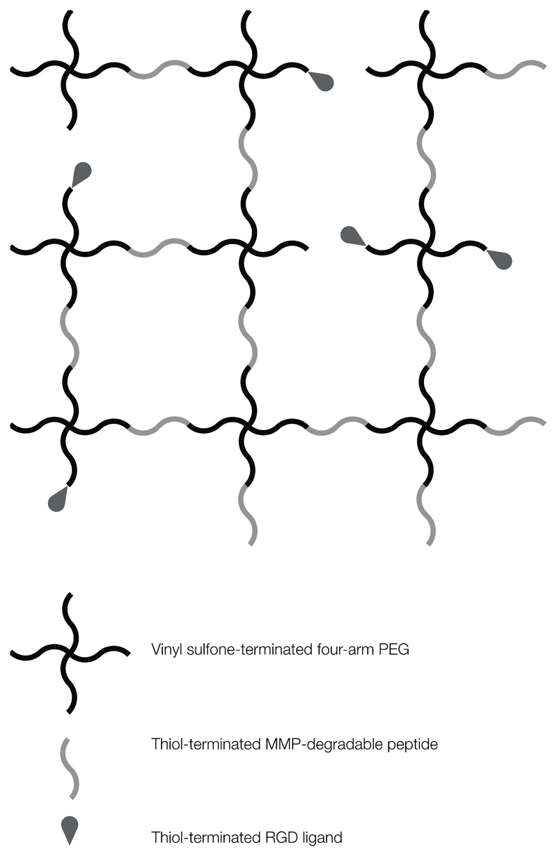
The role of soluble messengers in directing cellular behaviours has been recognized for decades. However, many cellular processes, including adhesion, migration and stem cell differentiation, are also governed by chemical and physical interactions with non-soluble components of the extracellular matrix (ECM). Among other effects, a cell's perception of nanoscale features such as substrate topography and ligand presentation, and its ability to deform the matrix via the generation of cytoskeletal tension play fundamental roles in these cellular processes. As a result, many biomaterials-based tissue engineering and regenerative medicine strategies aim to harness the cell's perception of substrate stiffness and nanoscale features to direct particular behaviours. However, since cell-ECM interactions vary considerably between two-dimensional (2-D) and three-dimensional (3-D) models, understanding their influence over normal and pathological cell responses in 3-D systems that better mimic the in vivo microenvironment is essential to translate such insights efficiently into medical therapies. This review summarizes the key findings in these areas and discusses how insights from 2-D biomaterials are being used to examine cellular behaviours in more complex 3-D hydrogel systems, in which not only matrix stiffness, but also degradability, plays an important role, and in which defining the nanoscale ligand presentation presents an additional challenge.

Keywords: Cell adhesion; Extracellular matrix; Hydrogel; Integrin; Stem cell.


Figures

Fig. 1
Fig. 1
Integrin-mediated cell-ECM adhesion. (a) Cells (yellow) residing on their ECM (black mesh). Red lines represent actin stress fibres and nuclei are shown in blue. (b) Integrin-mediated cell adhesion to the ECM requires clustering of multiple integrin receptors and FA complexes for efficient actin fibre assembly. (c) A detailed view of FA complexes connecting ECM-bound integrin receptors to the actin cytoskeleton. FA consist of proteins including, but not limited to vinculin, talin, paxillin and focal adhesion kinase.
Fig. 2
Fig. 2
Fluorescent micrographs of cells residing on different adhesive geometries (scale bar, 10 μm). Individual epithelial cells seeded on: (a) Y-shaped and (b) V-shaped adhesive fibronectin patterns on a non-adhesive background. Actin filaments (red) are wider and more numerous along non-adhesive edges, where they resist greater cytoskeletal tension. Vinculin (green) is concentrated at FA. Adapted with permission from ref. [62], © John Wiley & Sons, Inc. Individual MSC cultured on: (c) round and (d) stellar adhesive geometries of the same surface area, with stronger actin staining (red) and osteogenic differentiation associated with steeper angles. Adapted with permission from ref. [61], © Elsevier Ltd.
Fig. 3
Fig. 3
The effect of ligand spacing on FA clustering and actin cytoskeleton assembly in anchorage-dependent cells, shown to approximate scale. (a) A detailed view of integrin receptors bound to an Au-RGD-functionalised BCML substrate. Adhesion triggers recruitment of intracellular proteins, which aggregate to form FA. Lateral clustering of FA is followed by formation of actin fibres. (b) Substrates patterned with ligands spaced up to 58 nm apart enable FA formation and clustering, whereas (c) substrates with ligands spaced 73 nm or further apart do not support efficient FA formation, cell adhesion and spreading.
Fig. 4
Fig. 4
Schematic showing alginate hydrogels with varying ligand spacing and clustering. (a) RGD-functionalised alginate chains coil to form island amongst blank alginate coils. (b–c) Overall ligand density can be varied by altering island concentration, in turn varying ligand spacing. (d–e) Micro-nano-patterning of hydrogels using islands with different ligand numbers allows variation in ligand clustering while maintaining constant overall density.
Fig. 5
Fig. 5
Substrate stiffness influences cell morphology and differentiation. (a) Adherent cells (yellow) are unable to generate sufficient traction force to deform stiff hydrogel substrates (dark blue). As a result, they develop spread morphologies with many well-defined actin fibres (red). (b) By contrast, cells cultured on compliant substrates (pale blue) deform the matrix, and assume a more rounded morphology with fewer and less defined stress fibres. The distorted shapes of the hydrogel substrates of varying stiffness represent the deformation induced by the cell.
Fig. 6
Fig. 6
Microposts of varying height and stiffness induce different MSC morphologies. Micropost diameter 2 μm, spacing 4 μm, drawn to scale (scale bar 50 μm). (a) Short (0.97 μm), rigid microposts induce spread morphology and osteoblastic differentiation, whereas (b) MSC cultured on long (12.9 μm), flexible microposts favour adipogenic differentiation. (c) Schematic showing differences in flexibility of microposts subjected to equal forces, as a function of height.
Fig. 7
Fig. 7
Schematic of RGD-functionalised and MMP-degradable PEG hydrogel formed by click chemistry. Vinyl sulfone-terminated four-arm PEG is first functionalised with thiol-terminated RGD (in high stoichiometric deficit) and then cross-linked via thiol-terminated peptides containing MMP cleavage sites, forming an orthogonal but enzymatically degradable adhesive matrix.

References

    1. Wang N, Butler JP, Ingber DE. Mechanotransduction across the cell surface and through the cytoskeleton. Science. 1993;260:1124–7. - PubMed
    1. Murphy WL, McDevitt TC, Engler AJ. Materials as stem cell regulators. Nature Mater. 2014;13:547–57. - PMC - PubMed
    1. Arnold M, Cavalcanti-Adam EA, Glass R, Blümmel J, Eck W, Kantlehner M, et al. Activation of integrin function by nanopatterned adhesive interfaces. Chemphyschem. 2004;5:383–8. - PubMed
    1. Dalby MJ, Gadegaard N, Tare R, Andar A, Riehle MO, Herzyk P, et al. The control of human mesenchymal cell differentiation using nanoscale symmetry and disorder. Nature Mater. 2007;6:997–1003. - PubMed
    1. Mark von der K, Park J, Bauer S, Schmuki P. Nanoscale engineering of biomimetic surfaces: cues from the extracellular matrix. Cell Tissue Res. 2010;339:131–53. - PubMed

Publication types

Substances