. 2014 Nov 14;289(46):32253-32265.
doi: 10.1074/jbc.M114.578377. Epub 2014 Sep 29.

Hyaluronan synthase 2 protects skin fibroblasts against apoptosis induced by environmental stress

Affiliations

Hyaluronan synthase 2 protects skin fibroblasts against apoptosis induced by environmental stress

Yan Wang et al. J Biol Chem. .

Abstract

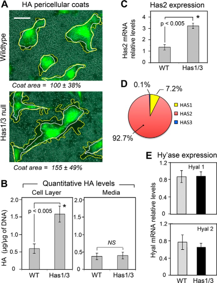
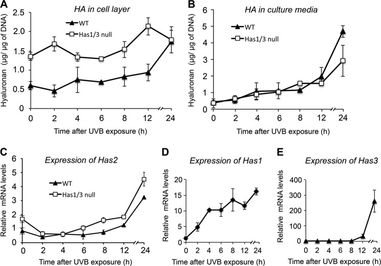
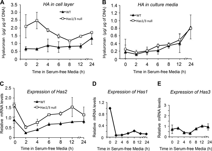
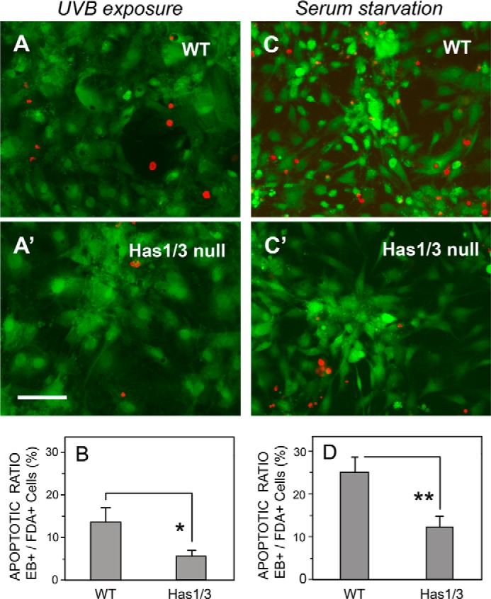
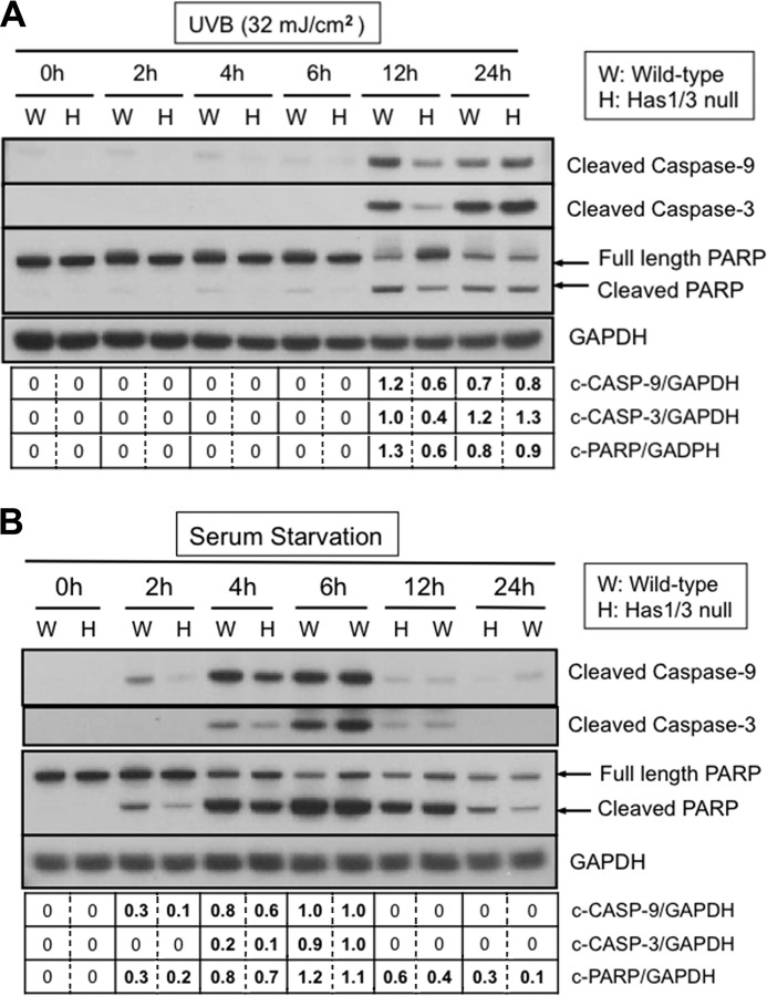
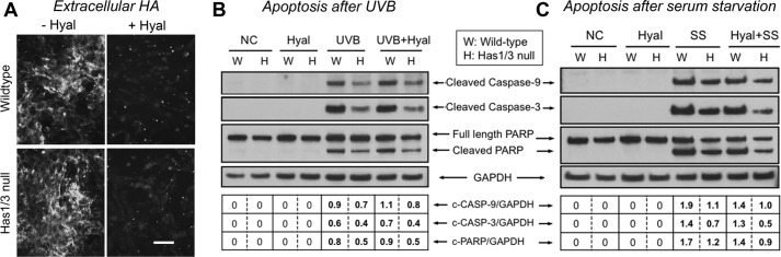
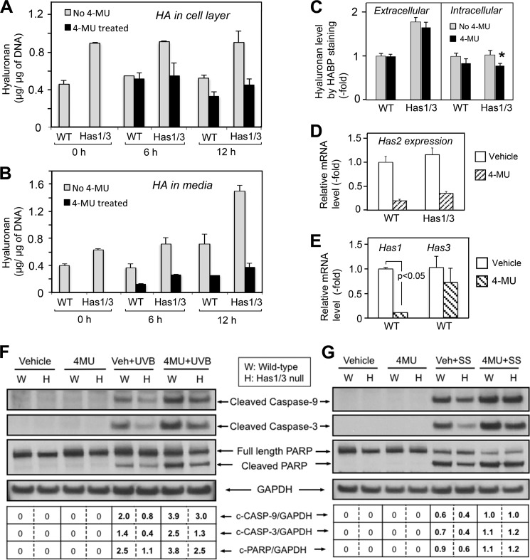
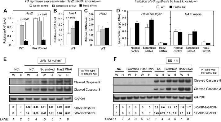
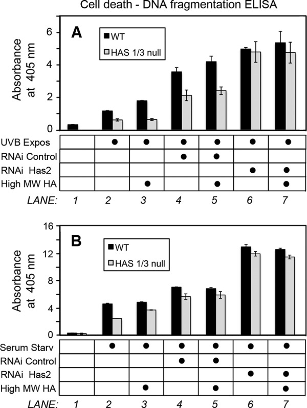
A balanced turnover of dermal fibroblasts is crucial for structural integrity and normal function of the skin. During recovery from environmental injury (such as UV exposure and physical wounding), apoptosis is an important mechanism regulating fibroblast turnover. We are interested in the role that hyaluronan (HA), an extracellular matrix molecule synthesized by HA synthase enzymes (Has), plays in regulating apoptosis in fibroblasts. We previously reported that Has1 and Has3 double knock-out (Has1/3 null) mice show accelerated wound closure and increased numbers of fibroblasts in the dermis. In the present study, we report that HA levels and Has2 mRNA expression are higher in cultured Has1/3 null primary skin fibroblasts than in wild type (WT) cells. Apoptosis induced by two different environmental stressors, UV exposure and serum starvation (SS), was reduced in the Has1/3 null cells. Hyaluronidase, added to cultures to remove extracellular HA, surprisingly had no effect upon apoptotic susceptibility to UVB or SS. However, cells treated with 4-methylumbelliferone to inhibit HA synthesis were sensitized to apoptosis induced by SS or UVB. When fibroblasts were transfected with Has2-specific siRNA that lowered Has2 mRNA and HA levels by 90%, both Has1/3 null and WT cells became significantly more sensitive to apoptosis. The exogenous addition of high molecular weight HA failed to reverse this effect. We conclude that Has1/3 null skin fibroblasts (which have higher levels of Has2 gene expression) are resistant to stress-induced apoptosis.

Keywords: Apoptosis; Caspase; Extracellular Matrix; Fibroblast; Hyaluronan; Skin; Stress.


Figures

FIGURE 1.
FIGURE 1.
Hyaluronan levels are increased and Has2 gene expression is up-regulated in Has1/3 null skin fibroblasts. A, living fibroblasts were labeled with calcein to visualize cell bodies, and the area occupied by the pericellular HA coat was assessed by the exclusion of RBCs from the cell perimeter. Yellow lines and white lines were artificially drawn to delineate the margin of the HA coat and cell membrane, respectively. Scale bar, 50 μm. Numbers below each panel are the mean value ± S.D. of three experiments (15 cells analyzed per experiment). B, the amount of HA derived from the cell layer (left) or media (right) of WT or Has1/3 null fibroblast cultures were analyzed by FACE and normalized by DNA content of each sample. Each data point is the mean ± S.E. (error bars) of four independent experiments. C, Has2 mRNA levels in WT and Has1/3 null cells, measured by qPCR. D, pie chart of relative mRNA levels of the Has isoforms in WT skin fibroblasts, measured by qPCR using the 2−ΔΔCt method and displayed as a percentage of total Has expression. E, hyaluronidase 1 and 2 (Hyal1 and Hyal2) mRNA levels in WT and Has1/3 null cells, as determined by qPCR. In C and E, each data point represents the mean ± S.E. of three independent experiments. *, significantly different from WT by Student's t test.
FIGURE 2.
FIGURE 2.
Changes in HA levels and Has mRNA levels in WT or Has1/3 null skin fibroblasts in response to UVB exposure. A and B, time courses are shown for HA levels in the cell layer (A) or media (B) of cultures after 32 mJ/cm2 UVB exposure, with quantification by FACE analysis. Each data point is the mean ± S.D. (error bars) of four independent experiments. Changes in gene expression of the HA synthases in WT and Has1/3 cells were determined by qPCR (mean ± half-range of two experiments) for Has2 (C), Has1 (D), and Has3 (E).
FIGURE 3.
FIGURE 3.
Changes in HA levels and Has mRNA levels in WT or Has1/3 null skin fibroblasts in response to serum starvation. Time courses of HA levels in the cell layer (A) or media (B) during 24-h serum starvation are shown, measured by FACE analysis; each data point is the mean ± S.D. (error bars) of four experiments. Changes in gene expression of the HA synthases during serum starvation of WT and Has1/3 cells were determined by qPCR (mean ± half-range of two experiments) for Has2 (C), Has1 (D), and Has3 (E).
FIGURE 4.
FIGURE 4.
Has1/3 null fibroblasts are relatively resistant to apoptosis. Panels on left, FDA/EB “live/dead” staining of cells 12 h after UVB irradiation (32 mJ/cm2). Representative fluorescent microscopy images of WT cells (A) and Has1/3 null cells (A′) are shown above; quantitation of the ratio of dead (EB+) to live (FDA+) cells is shown below in (B). Panels on right, FDA/EB staining of cells after 4 h of serum starvation. Representative images (C and C′) and quantitation of the EB/FDA ratio (graph, D) are shown. Scale bar, 100 μm. Each bar in the graphs is the mean ± S.D. (error bars) of three independent experiments. Comparison was by Student's t test: *, p < 0.025; **, p < 0.001.
FIGURE 5.
FIGURE 5.
Attenuated apoptotic responses in Has1/3 null fibroblasts involve reduced caspase activation. A, representative Western blots of cleaved caspase-9, cleaved caspase-3, and full-length and cleaved PARP in skin fibroblasts at different times after UVB exposure (32 mJ/cm2). GAPDH was used as a loading control. B, Western blots as in A, from fibroblasts harvested at different times after serum starvation. Data in the table below these blots are the mean from densitometric scans in the corresponding lane, averaged from two independent experiments; the variance was less than 20% of the mean in all cases.
FIGURE 6.
FIGURE 6.
Apoptosis resistance of skin fibroblasts is not mediated by HA in the extracellular matrix. A, extracellular HA in skin fibroblast cultures treated with S. dysgalactiae hyaluronidase (0.3 unit/ml, 10 min at 37 °C) (+Hyal) or in untreated control cultures (−Hyal) and then immunostained with bHABP/streptavidin-Alexa Fluor 488. Scale bar, 100 μm. B, representative Western blots of cleaved caspase-9 and -3 and cleaved PARP in skin fibroblasts treated with either the hyaluronidase (Hyal) or UVB irradiation (UVB) or both (UVB + Hyal), versus untreated normal controls (NC). C, Western blots of cleaved caspase-9 and -3 and cleaved PARP in skin fibroblasts treated with either Hyal or serum starvation (SS) or both (Hyal + SS), versus untreated normal controls (NC). GAPDH was examined as a loading control.
FIGURE 7.
FIGURE 7.
4-MU treatment sensitizes skin fibroblasts to apoptosis. A, HA levels in the cell layer (A) or culture media (B) of fibroblasts treated with 4-MU (1 mm) for either 6 or 12 h were quantified by FACE analysis. C, quantitation of fluorescence intensity of extracellular HA (left) and intracellular HA (right) staining with bHABP, in skin fibroblasts treated with 4-MU for 6 h and analyzed by image processing. Gray bars, no 4-MU; black bars, +4-MU. *, p < 0.05 by Student's t test (significant difference from vehicle-treated cells). Changes in Has2 mRNA levels in both types of fibroblasts (D) or in Has1 and Has3 mRNA levels in WT fibroblasts (E), treated with 4-MU (1 mm) or vehicle alone for 6 h, were analyzed by qPCR. Each data point is the mean ± S.D. of two independent experiments. F, representative Western blots of cleaved caspase-9 and -3 and cleaved PARP in skin fibroblasts treated with 4-MU or vehicle control for 6 h, followed by 32 mJ/cm2 UVB (4-MU + UVB and Veh + UVB). G, Western blots of cleaved caspase-9 and -3 and cleaved PARP in skin fibroblasts treated with either 4-MU or vehicle control for 6 h followed by 4 h of serum starvation (4-MU + SS and Veh + SS). GAPDH, loading control.
FIGURE 8.
FIGURE 8.
Knockdown of Has2 gene expression by Has2 siRNA transfection sensitizes skin fibroblasts to apoptosis. mRNA expression levels of Has2 (A), Has1 (B), and Has3 (C) in either WT or Has1/3 null fibroblasts, analyzed by qPCR. No Rx control, untreated normal control; Scrambled siRNA, transfected with non-targeted scrambled siRNA; Has2 siRNA, transfected with Has2 siRNA. Each bar represents the mean ± S.D. (error bars) of two independent experiments. D, HA levels in the cell layer (left) or media (right) from fibroblast cultures, treated as described for A–C and analyzed by FACE. Each bar is the mean ± S.D. of two independent experiments. E and F, Western blots of skin fibroblasts after UVB (32 mJ/cm2) or serum starvation (4 h), respectively, showing effects upon the expression of cleaved caspase-9 and -3, relative to GAPDH loading controls, in cells transfected with siRNA against Has2 (Has2 siRNA), non-targeted scrambled siRNA (Scrambled), or untreated normal control (NC).
FIGURE 9.
FIGURE 9.
External addition of high molecular weight HA fails to rescue the apoptotic sensitization caused by Has2 RNAi. Shown is cell death/DNA fragmentation in fibroblasts transfected with Has2 siRNA (RNAi Has2) or with scrambled siRNA (RNAi Control), prior to a UVB 32 mJ/cm2 exposure (A) or 4-h serum starvation (B), in the presence or absence of high molecular weight HA (High MW HA). Apoptosis was measured with a cell death detection ELISA kit.

References

    1. Kubo K., Kuroyanagi Y. (2005) A study of cytokines released from fibroblasts in cultured dermal substitute. Artif. Organs 29, 845–849 - PubMed
    1. Wong T., McGrath J. A., Navsaria H. (2007) The role of fibroblasts in tissue engineering and regeneration. Br. J. Dermatol. 156, 1149–1155 - PubMed
    1. Werner S., Krieg T., Smola H. (2007) Keratinocyte-fibroblast interactions in wound healing. J. Invest. Dermatol. 127, 998–1008 - PubMed
    1. Jelaska A., Korn J. H. (2000) Role of apoptosis and transforming growth factor β1 in fibroblast selection and activation in systemic sclerosis. Arthritis Rheum. 43, 2230–2239 - PubMed
    1. Jun J. B., Kuechle M., Min J., Shim S. C., Kim G., Montenegro V., Korn J. H., Elkon K. B. (2005) Scleroderma fibroblasts demonstrate enhanced activation of Akt (protein kinase B) in situ. J. Invest. Dermatol. 124, 298–303 - PubMed

Publication types

LinkOut - more resources