. 2014 Sep 29;206(7):877-94.
doi: 10.1083/jcb.201401146.

Polo-like kinase 3 regulates CtIP during DNA double-strand break repair in G1

Affiliations

Polo-like kinase 3 regulates CtIP during DNA double-strand break repair in G1

Olivia Barton et al. J Cell Biol. .

Abstract

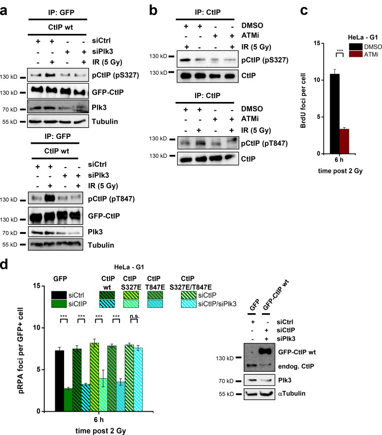
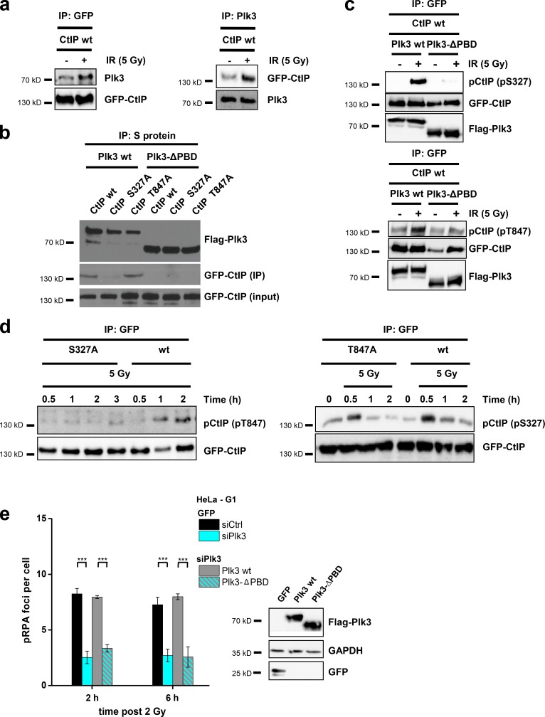
DNA double-strand breaks (DSBs) are repaired by nonhomologous end joining (NHEJ) or homologous recombination (HR). The C terminal binding protein-interacting protein (CtIP) is phosphorylated in G2 by cyclin-dependent kinases to initiate resection and promote HR. CtIP also exerts functions during NHEJ, although the mechanism phosphorylating CtIP in G1 is unknown. In this paper, we identify Plk3 (Polo-like kinase 3) as a novel DSB response factor that phosphorylates CtIP in G1 in a damage-inducible manner and impacts on various cellular processes in G1. First, Plk3 and CtIP enhance the formation of ionizing radiation-induced translocations; second, they promote large-scale genomic deletions from restriction enzyme-induced DSBs; third, they are required for resection and repair of complex DSBs; and finally, they regulate alternative NHEJ processes in Ku(-/-) mutants. We show that mutating CtIP at S327 or T847 to nonphosphorylatable alanine phenocopies Plk3 or CtIP loss. Plk3 binds to CtIP phosphorylated at S327 via its Polo box domains, which is necessary for robust damage-induced CtIP phosphorylation at S327 and subsequent CtIP phosphorylation at T847.


Figures

Figure 1.
Figure 1.
CtIP enhances translocation formation in G1. (a) Identification of cell cycle phases (see first paragraph of the Results for description). arb. units, arbitrary units. (b) γ-H2AX foci in G1-phase HeLa cells after CtIP siRNA. (c) PCC spread from G1-phase 82-6 fibroblasts fused with mitotic HeLa cells. FISH staining was performed with chromosome probes 1, 2, and 4. The one-chromatid G1 PCC spread is encircled with a dashed line; the two-chromatid mitotic HeLa cell is outside this area. (d) Chromosome breaks in G1-phase 82-6 fibroblasts after CtIP siRNA. The image shows a chromosome break indicated by the presence of an additional fragment of the stained chromosome. (e) Translocations in G1-phase 82-6 fibroblasts after CtIP siRNA. The image shows a translocation indicated by the color junction (±SEM from at least three experiments). siCtrl, control siRNA; siCtIP, CtIP siRNA. *, P < 0.05.
Figure 2.
Figure 2.
RPA loading and phosphorylation occurs in G1 at complex DSBs. (a) Detection of cytosolic and chromatin-bound CtIP, RPA2, and Rad51 in proliferating (prol.) and confluency-arrested (G0) 82-6 fibroblasts 4 h after IR. The Rad51 signals are shown in two intensity settings (Rad51*, stronger intensity setting). GAPDH and H3 were used as markers for the cytosolic and chromatin-bound fraction, respectively. (b) Time course for the formation and disappearance of pRPA2 in whole cell extracts from confluency-arrested 82-6 fibroblasts. Caspase7, which provides a pronounced signal in cells treated with the apoptosis induction factor TNF and cycloheximide, was used to control for apoptosis. 1 µM camptothecin (CPT) treatment (for 1 h) was used to control for the presence of S-phase cells. (c and d) Detection of pRPA (c) and BrdU (d) foci in G1-phase HeLa cells treated with CtIP siRNA and irradiated with 2 Gy α particles. The mean number of foci per G1 cell (middle) and the fraction of G1 cells with more than five foci (right) are shown (±SEM from at least three experiments). siCtrl, control siRNA; siCtIP, CtIP siRNA. ***, P < 0.001.
Figure 3.
Figure 3.
CtIP function in G1 requires S327 and T847 phosphorylation. (a) pRPA foci in GFP-positive (GFP+), G1-phase HeLa cells irradiated with 2 Gy α particles. Endogenous CtIP was depleted by siRNA, and cells were transfected with various GFP-CtIP plasmids. Knockdown efficiency for CtIP and transfection efficiencies for GFP-CtIP were confirmed by Western blotting. endog., endogenous. (b) Insect cell–purified CtIP-wt, CtIP-S327A, and CtIP-T847A proteins were incubated with recombinant Cdk2/CyclinA and analyzed with phosphospecific antibodies for CtIP-pS327 or CtIP-pT847. (c) Phosphorylation of CtIP in vivo. HEK293T cells were treated with CtIP siRNA, transfected with GFP-CtIP-wt, GFP-CtIP-S327A, or GFP-CtIP-T847A plasmids, enriched in G1 (≥90%), and irradiated with 5 Gy. GFP-CtIP was obtained by IP and analyzed for pS327 or pT847 by immunoblotting. (d) Analysis of excision events in GC92 cells containing an NHEJ reporter substrate. Excision formation from the repair of two I-SceI–induced DSBs results in a CD4-positive signal. Cells were treated with CtIP siRNA and transfected with various RFP-CtIP plasmids. Excisions were measured by the fraction of RFP-positive cells that exhibited a CD4-positive signal relative to all RFP-positive cells. Results were normalized to control siRNA-treated cells that were transfected with an RFP empty vector. Knockdown efficiencies for CtIP and transfection efficiencies for RFP-CtIP plasmids were confirmed by Western blotting (±SEM from at least three experiments). siCtrl, control siRNA; siCtIP, CtIP siRNA. ***, P < 0.001.
Figure 4.
Figure 4.
Plk3 is required for resection in G1 and phosphorylates CtIP in vitro. (a) pRPA (left) and BrdU (middle) foci in G1-phase HeLa cells treated with Plk3 siRNA or Plki and irradiated with 2 Gy α particles. Knockdown efficiency for Plk3 was confirmed by Western blotting. Detection of pRPA2 at 4 h after IR or CPT (1 µM for 1 h) in whole cell extracts from confluency-arrested 82-6 fibroblasts with and without Plki (right). GAPDH was used as a loading control, and CyclinA was used as a marker for S/G2 cells. (b) Endogenous CtIP obtained by IP of CtIP from untransfected HeLa cells or GFP-CtIP obtained by IP of GFP from transfected HeLa cells was incubated in vitro with recombinant, constitutively active Plk3. Phosphorylation was measured by 32P incorporation. The bands representing pCtIP or GFP-pCtIP are indicated. (c) Insect cell purified CtIP-wt protein was incubated in vitro with Plk3 or Cdk2/CyclinA and analyzed with phosphospecific antibodies for CtIP-pT847 or CtIP-pS327. (d) Insect cell purified CtIP-wt and CtIP-S327A/T847A proteins were incubated in vitro with Plk3 and analyzed with an antibody against CtIP (±SEM from at least three experiments). siCtrl, control siRNA; siPlk3, Plk3 siRNA. ***, P < 0.001.
Figure 5.
Figure 5.
Plk3 phosphorylates CtIP in vivo. (a) HEK293T cells were treated with Plk3 and CtIP siRNAs, transfected with GFP-CtIP plasmids, enriched in G1 (≥90%), and irradiated with 5 Gy. GFP-CtIP was obtained by IP and analyzed for pS327 at 30 min after IR or pT847 at 2 h after IR by immunoblotting. (b) Confluent 82-6 fibroblasts were treated with ATMi and irradiated with 5 Gy. Endogenous CtIP was obtained by IP and analyzed for pS327 at 30 min after IR or pT847 at 2 h after IR by immunoblotting. (c) BrdU foci in ATMi-treated, G1-phase HeLa cells irradiated with 2 Gy α particles. (d) pRPA foci in GFP-positive (GFP+), G1-phase HeLa cells after 2 Gy α-particle irradiation. Cells were treated with CtIP and Plk3 siRNAs and transfected with GFP-CtIP plasmids. Knockdown efficiencies for CtIP and Plk3 as well as transfection efficiencies for GFP-CtIP were confirmed by Western blotting (±SEM from three experiments). endog., endogenous; siCtrl, control siRNA; siCtIP, CtIP siRNA; siPlk3, Plk3 siRNA. ***, P < 0.001.
Figure 6.
Figure 6.
Plk3 interacts with CtIP to promote resection in G1. (a) Interaction of GFP-CtIP and Plk3 after 5 Gy IR. HEK293T cells were treated with CtIP siRNA and transfected with a GFP-CtIP plasmid. GFP-CtIP or Plk3 were obtained by IP, and the precipitates were analyzed by immunoblotting. (b) Interaction of Plk3-ΔPBD with CtIP-S327A and CtIP-T847A at 30 min after 5 Gy. HEK293T cells were treated with CtIP and Plk3 siRNAs and transfected with GFP-CtIP and SFB-Plk3 plasmids. Plk3 was obtained by IP, and the precipitates were analyzed by immunoblotting. (c) CtIP phosphorylation in a Plk3-ΔPBD mutant in vivo. HEK293T cells were treated with CtIP and Plk3 siRNAs, transfected with GFP-CtIP and SFB-Plk3 plasmids, and irradiated with 5 Gy. GFP-CtIP was obtained by IP and analyzed for pS327 at 30 min after IR or pT847 at 2 h after IR by immunoblotting. (d) HEK293T cells were treated with CtIP siRNA, transfected with GFP-CtIP plasmids, and irradiated with 5 Gy. GFP-CtIP was obtained by IP and analyzed for pS327 or pT847 by immunoblotting. (e) pRPA foci in Flag-positive (Flag+), G1-phase HeLa cells after 2 Gy α-particle irradiation. Cells were treated with Plk3 siRNA and transfected with SFB-Plk3 plasmids. Transfection efficiencies were confirmed by Western blotting (±SEM from three experiments). siCtrl, control siRNA; siPlk3, Plk3 siRNA. ***, P < 0.001.
Figure 7.
Figure 7.
Plk3 enhances the formation of translocations and genomic rearrangements in G1. (a) γ-H2AX foci in G1-phase 82-6 fibroblasts treated with Plk3 siRNA or Plki. Plk3 knockdown efficiency was confirmed by Western blotting. (b) Chromosome breaks in G1-phase 82-6 fibroblasts treated with Plki. (c) Translocations in G1-phase 82-6 fibroblasts treated with Plk3 siRNA or Plki. (d) Analysis of excision events in GC92 cells. Cells were treated with CtIP and/or Plk3 siRNA, and excisions were measured by the fraction of cells that exhibited a CD4-positive signal relative to all cells. Results were normalized to control siRNA-treated cells. The knockdown efficiencies for CtIP and Plk3 were confirmed by Western blotting (±SEM from at least three experiments). siCtrl, control siRNA; siCtIP, CtIP siRNA; siPlk3, Plk3 siRNA. *, P < 0.05; ***, P < 0.001.
Figure 8.
Figure 8.
Plk3 is required for the repair of complex DSBs and DSBs in G1-phase Ku−/− MEFs. (a) γ-H2AX foci in G1-phase 82-6 fibroblasts treated with CtIP siRNA and/or Plki and irradiated with 2 Gy α particles. (b) γ-H2AX foci in G1-phase wt and Ku80−/− MEFs treated with Lig1/3 siRNA and/or PARP inhibition (PARPi). Knockdown efficiencies were confirmed by Western blotting. (c) γ-H2AX foci in G1-phase wt and Ku80−/− MEFs treated with CtIP siRNA and/or Plki and PARP inhibition. (d) Survival of G1-phase wt and Ku80−/− MEFs after Plki. G1 cells were obtained by mitotic shake-off followed by 2-h incubation before IR (±SEM from at least three experiments; Jackman and O’Connor, 2001). siCtrl, control siRNA; siCtIP, CtIP siRNA; siLig1/3, Lig1/3 siRNA. *, P < 0.05; ***, P < 0.001.

References

    1. Anantha, R.W., Vassin V.M., and Borowiec J.A.. 2007. Sequential and synergistic modification of human RPA stimulates chromosomal DNA repair. J. Biol. Chem. 282:35910–35923 10.1074/jbc.M704645200 - DOI - PubMed
    1. Anger, M., Kues W.A., Klima J., Mielenz M., Kubelka M., Motlik J., Esner M., Dvorak P., Carnwath J.W., and Niemann H.. 2003. Cell cycle dependent expression of Plk1 in synchronized porcine fetal fibroblasts. Mol. Reprod. Dev. 65:245–253 10.1002/mrd.10289 - DOI - PubMed
    1. Bahassi, M., Conn C.W., Myer D.L., Hennigan R.F., McGowan C.H., Sanchez Y., and Stambrook P.J.. 2002. Mammalian Polo-like kinase 3 (Plk3) is a multifunctional protein involved in stress response pathways. Oncogene. 21:6633–6640 10.1038/sj.onc.1205850 - DOI - PubMed
    1. Beausoleil, S.A., Villén J., Gerber S.A., Rush J., and Gygi S.P.. 2006. A probability-based approach for high-throughput protein phosphorylation analysis and site localization. Nat. Biotechnol. 24:1285–1292 10.1038/nbt1240 - DOI - PubMed
    1. Beucher, A., Birraux J., Tchouandong L., Barton O., Shibata A., Conrad S., Goodarzi A.A., Krempler A., Jeggo P.A., and Löbrich M.. 2009. ATM and Artemis promote homologous recombination of radiation-induced DNA double-strand breaks in G2. EMBO J. 28:3413–3427 10.1038/emboj.2009.276 - DOI - PMC - PubMed

Publication types

MeSH terms