

doi: 10.1021/nl502385k.
Epub 2014 Sep 30.
Multifunctional RNA nanoparticles
Affiliations
- PMID: 25267559
- PMCID: PMC4189619
- DOI: 10.1021/nl502385k
Item in Clipboard
Multifunctional RNA nanoparticles
Nano Lett.
.
Abstract
Our recent advancements in RNA nanotechnology introduced novel nanoscaffolds (nanorings); however, the potential of their use for biomedical applications was never fully revealed. As presented here, besides functionalization with multiple different short interfering RNAs for combinatorial RNA interference (e.g., against multiple HIV-1 genes), nanorings also allow simultaneous embedment of assorted RNA aptamers, fluorescent dyes, proteins, as well as recently developed RNA-DNA hybrids aimed to conditionally activate multiple split functionalities inside cells.
Keywords: RNA interference; RNA nanoparticles; RNA nanotechnology; RNA−DNA hybrid reassociation; aptamers.
Figures
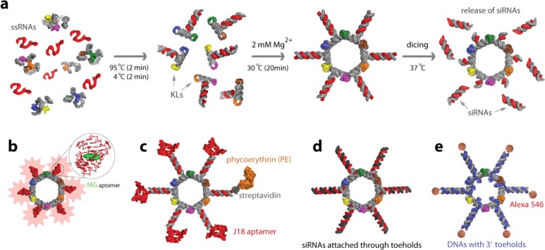
Schematic representation
of assemblies leading to the formation
of RNA nanorings functionalized with (a) Dicer substrate RNAs, (b)
malachite green (MG) aptamers for in vitro visualization, (c) J18
aptamers for cell targeting and phycoerythrin for visualization in
vivo, (d) Dicer substrate RNAs introduced via the toehold interactions,
and (e) RNA–DNA hybrids with split functionalities (RNAi and
FRET). Functional siRNAs can be released by Dicer nuclease. KLs stand
for kissing loops.
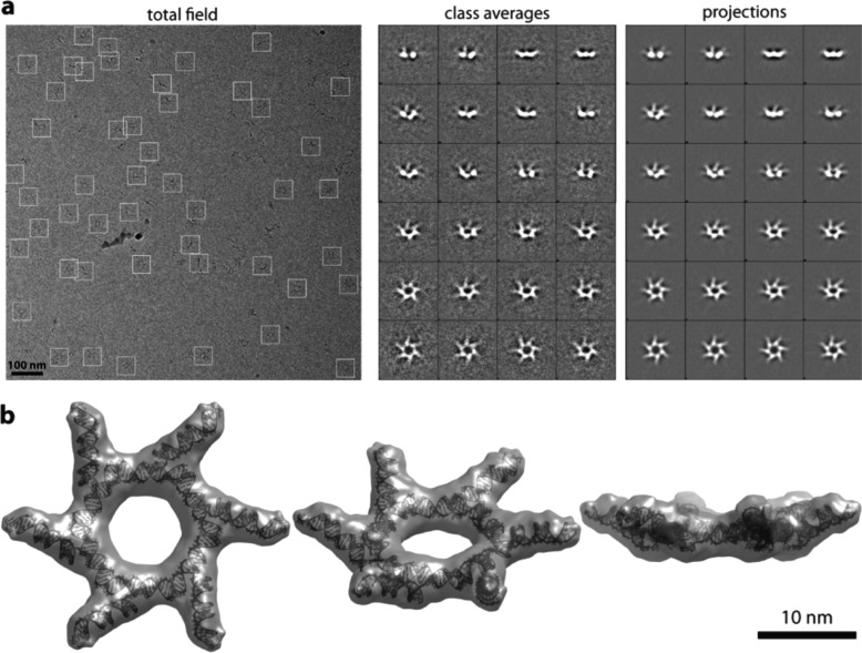
Structural characterization by cryo-EM
of RNA nanorings functionalized
with six DS RNAs. (a) A typical cryo-EM image of the DS RNA nanoring
particles (left panel). Class averages for each DS RNA nanoring as
observed by cryo-EM (central panel), with corresponding projections
of the reconstructed three-dimensional structure (right panel). (b)
Single particle reconstruction of functionalized RNA nanorings. Different
views of the model fit with the electron density volume are shown.
The volume map is thresholded at the minimum level at which all the
atoms of the model can be fit inside the volume. The resolution is
16 Å.
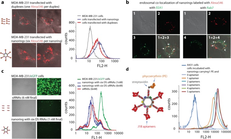
Cell uptake,
endosomal colocalization, silencing, and RNA aptamer
mediated binding efficiencies of functional nanorings. (a) Transfection
efficiencies using human breast cancer cells (MDA-MB-231). DS RNAs
(60 nM final) covalently labeled with one Alexa 546 per duplex were
compared to the functionalized nanorings (10 nM final) labeled with
six Alexa 546 dyes. One day after the transfection, the efficiencies
were analyzed by confocal fluorescence microscopy and flow cytometry
experiments. (b) Studying the localization of nanorings with commonly
used markers for endosomal compartments Early Endosome Antigen 1 (EEA1)
and Rab7. (c) GFP knockdown assays using human breast cancer cells
(MDA-MB-231/GFP) which stably express enhanced GFP (eGFP). Fluorescence
microscopy (left panel) and statistical analysis (30000 cells per
sample) of flow cytometry experiments (right panel) of eGFP expression
3 days after the transfection of cells with DS RNA duplexes and nanorings
functionalized with six DS RNAs against eGFP. The ratio of DS RNA
duplexes to DS RNA functionalized nanorings was 6:1. (d) Nanorings
labeled with phycoerythrin (PE) and containing different numbers of
the EGFR-specific J18 aptamer selected to specifically bind EGFR expressed
on A431 cells were tested for relative binding efficiencies using
FACS. The J18 RNA aptamer model is a conceptual cartoon, based on
the minimum free energy secondary structure (MFE). Image numbers in
(b) correspond to differential interference contrast (DIC) images
(1), Alexa546 emission (2), EAA1 antibody staining (3), and Rab7 antibody
staining (4). Images (1 + 2 + 3) and (1 + 2 + 4) are superpositions
of three different images.
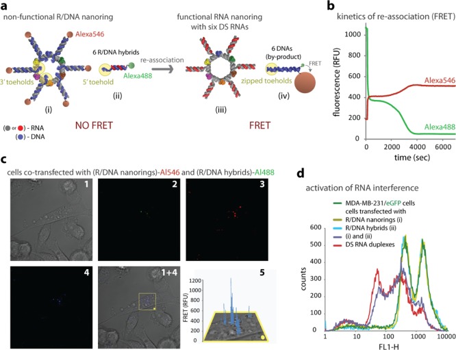
Activation
of different functionalities by RNA–DNA hybrids.
(a) Scheme showing an activation of multiple functionalities (RNAi,
FRET) upon reassociation of nonfunctional nanorings decorated with
RNA–DNA hybrids and six nonfunctional cognate RNA–DNA
hybrids. (b) FRET time traces during reassociation of hybrid nanorings
labeled with Alexa546 and cognate hybrids labeled with Alexa488. (c)
Intracellular FRET experiments: cells were cotransfected with hybrid
nanorings and cognate hybrids labeled with Alexa546 and Alexa488,
respectively. Images were taken the next day. (d) GFP knockdown assays.
Three days after transfection of MDA-MB-231/GFP cells with hybrid
nanorings and cognate hybrids programmed to release DS RNAs, eGFP
expression was statistically analyzed with flow cytometry experiments.
As the control, DS RNA duplexes against eGFP were used. Please note
that individually neither hybid nanorings nor hybrids cause decrease
in eGFP production. Image numbers in (c) correspond to differential
interference contrast (DIC) images (1), Alexa488 emission (2), Alexa546
emission (3), bleed-through corrected FRET image (4), and 3D chart
representation of zoomed fragment indicated by a yellow box of bleed-through
corrected FRET image with the yellow dot indicating the correspondence
(5).
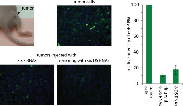
In vivo studies of nanorings functionalized with six DS RNAs in
a tumor xenograft mouse model. Fluorescent imaging of tumors and corresponding
quantification after 5 days postinjections in vivo demonstrate significant
levels of eGFP silencing caused by nanorings functionalized with six
DS RNAs compared to free siRNAs. Free DS RNAs were used at six times
higher concentrations. Error bars denote ±SEM; N = 2.
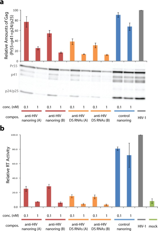
HIV-1 expression and production is inhibited by functional nanorings.
(a) HIV-1 expression inside the cell was measured at 48 h post-transfection.
HeLa cells were lysed and probed by Western blotting for HIV-1 proteins.
Positions of Pr55Gag (Pr55), matrix-capsid (p41), and capsid/capsid-SP1
(p24/p25) are indicated. Quantification of total cell-associated Gag:
Pr55 + p41 + p25 + p24. Total Gag in virus control (HIV-1) without
nanorings or Dicer substrate (DS) RNAs set at 100. Error bars denote
±SEM; N = 4. (b) HeLa cells were transfected
with pNL4-3 (full-length HIV-1 molecular clone), with and without
nanorings or DS RNAs. Virus supernatant was harvested 48 h post-transfection,
and the reverse transcriptase (RT) production was measured (this assay
quantifies the amounts of virus produced by the cells); data are shown
normalized to virus controls (HIV-1) without functional nanorings
or DS RNAs. Mock represents untrasfected HeLa cells. Corresponding
mixtures of six different anti-HIV DS RNAs (A and B) were used as
positive controls. Nanoring control without any anti-HIV DS RNAs was
used as a negative control. Error bars denote ±SEM; N = 4.
References
Publication types
MeSH terms
Substances
Grants and funding
LinkOut - more resources
Full Text Sources
Other Literature Sources
Research Materials
