. 2014 Oct 14;111(41):14852-7.
doi: 10.1073/pnas.1403271111. Epub 2014 Sep 29.

Antigen affinity and antigen dose exert distinct influences on CD4 T-cell differentiation

Affiliations

Antigen affinity and antigen dose exert distinct influences on CD4 T-cell differentiation

Simone Keck et al. Proc Natl Acad Sci U S A. .

Abstract

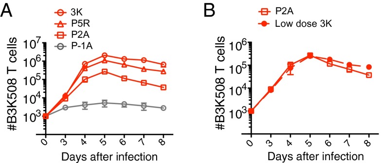
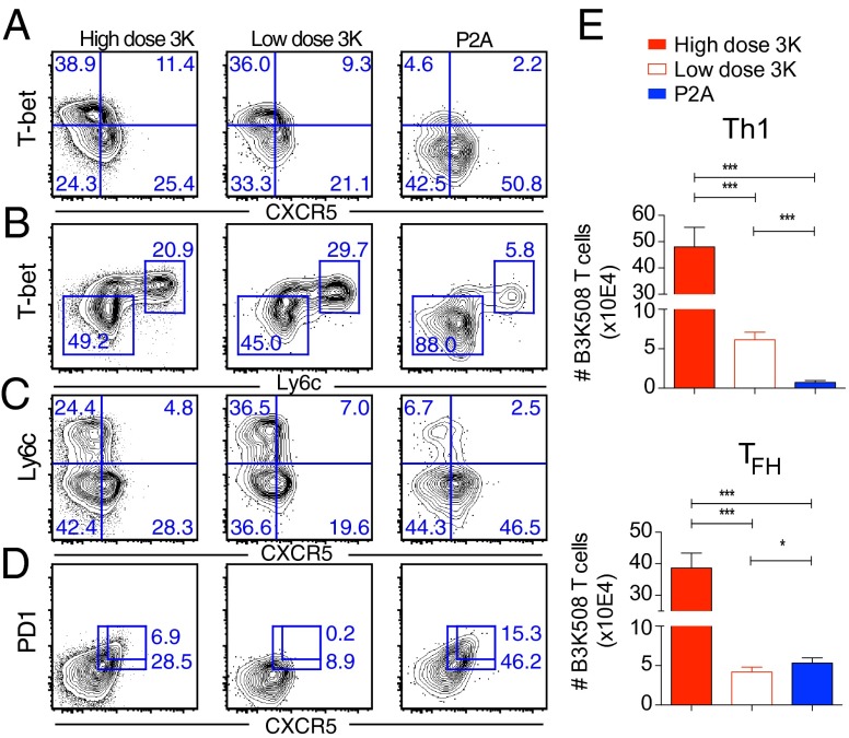
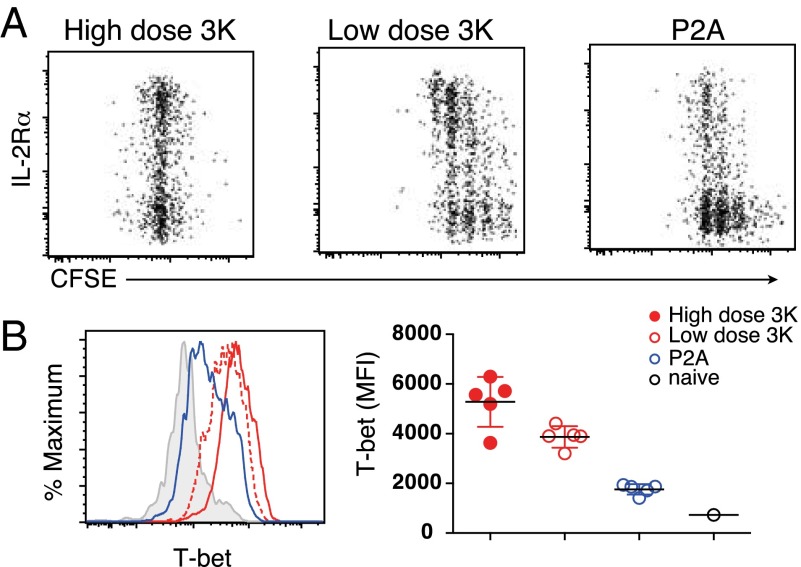
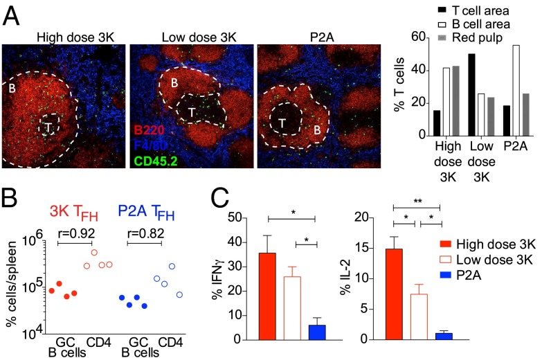
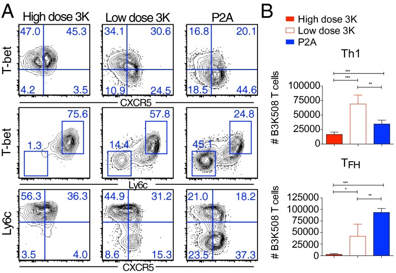
Cumulative T-cell receptor signal strength and ensuing T-cell responses are affected by both antigen affinity and antigen dose. Here we examined the distinct contributions of these parameters to CD4 T-cell differentiation during infection. We found that high antigen affinity positively correlates with T helper (Th)1 differentiation at both high and low doses of antigen. In contrast, follicular helper T cell (TFH) effectors are generated after priming with high, intermediate, and low affinity ligand. Unexpectedly, memory T cells generated after priming with very low affinity antigen remain impaired in their ability to generate secondary Th1 effectors, despite being recalled with high affinity antigen. These data challenge the view that only strongly stimulated CD4 T cells are capable of differentiating into the TFH and memory T-cell compartments and reveal that differential strength of stimulation during primary T-cell activation imprints unique and long lasting T-cell differentiation programs.

Keywords: follicular helper; infection; lymphocytes.


Conflict of interest statement

The authors declare no conflict of interest.

Figures

Fig. 1.
Fig. 1.
Establishing the TCR affinity hierarchy. (A and B) Ten thousand naïve B3K508 T cells were transferred into CD45.1 congenic mice followed by infection with Lm.3K or Lm.variant strains. Low dose Lm.3K infection in B corresponds to 105 cfu. Mean number of B3K508 T cells recovered from spleen and lymph nodes over the first 8 d of infection. Data represent n ≥ 3 for each data point and are representative of two independent experiments.
Fig. 2.
Fig. 2.
Antigen affinity and effector T-cell differentiation. Ten thousand B3K508 T cells were transferred into congenic mice followed by infection with Lm.3K or Lm.P2A. (AD) Representative flow cytometry plots of B3K508 T cells at day 6. (E) Absolute number of Th1 (T-bethighCXCR5) and TFH (T-betlowCXCR5+) effectors recovered from spleen and lymph nodes at day 6. Data represent n ≥ 3 and are representative of three independent experiments. (*P < 0.05, ***P < 0.0001).
Fig. 3.
Fig. 3.
Antigen affinity and early IL-2 induction. Two hundred thousand carboxyfluorescein succinimidyl ester (CFSE)-labeled B3K508 T cells were transferred into congenic mice followed by infection with Lm.3K or Lm.P2A. CD25 proliferation (A) and T-bet expression (B) were assessed by flow cytometry after 2 (A) and 3 (B) d. T-bet MFI is shown for T cells that have undergone at least one division. Data represent n = 3 for each data point and are representative of two independent experiments. (**P < 0.001, ***P < 0.0001).
Fig. 4.
Fig. 4.
Function and location of low affinity activated T cells. (A) Splenic sections were stained for B220 (red) to identify B-cell zones, F4/80 (blue) to identify red pulp area, and CD45.2 (green) to identify B3K508 donor T cells. Percentage of B3K508 T cells located in the indicated areas at day 6. (B) Th1 and TFH cells were sorted from infected mice and transferred into CD3ε−/− mice immunized 1 d earlier with 3K-OVA and alum. GC B-cell numbers correlate with TFH cell numbers. (C) Intracellular cytokine production was assessed by flow cytometry. Data represent n ≥ 3 mice per group and at least two independent experiments.
Fig. 5.
Fig. 5.
Low affinity TCR–pMHC ligands support memory T-cell differentiation. Phenotype (A) and absolute cell number (B) of B3K508 T cells recovered at day 4 after recall infection with virulent Lm.3K. Data represent n ≥ 3 and are representative of three independent experiments. (*P < 0.05, ***P < 0.0001).
Fig. 6.
Fig. 6.
Effector and memory T-cell differentiation in B-cell–deficient mice. Phenotype of B3K508 T cells recovered from spleen and lymph nodes of B6 or mb-1cre mice at day 6 after infection. Data representative of two independent experiments.

References

    1. Crotty S. Follicular helper CD4 T cells (TFH) Annu Rev Immunol. 2011;29:621–663. - PubMed
    1. Choi YS, et al. ICOS receptor instructs T follicular helper cell versus effector cell differentiation via induction of the transcriptional repressor Bcl6. Immunity. 2011;34(6):932–946. - PMC - PubMed
    1. Pepper M, Pagán AJ, Igyártó BZ, Taylor JJ, Jenkins MK. Opposing signals from the Bcl6 transcription factor and the interleukin-2 receptor generate T helper 1 central and effector memory cells. Immunity. 2011;35(4):583–595. - PMC - PubMed
    1. Whitmire JK, et al. Requirement of B cells for generating CD4+ T cell memory. J Immunol. 2009;182(4):1868–1876. - PMC - PubMed
    1. Corbin GA, Harty JT. Duration of infection and antigen display have minimal influence on the kinetics of the CD4+ T cell response to Listeria monocytogenes infection. J Immunol. 2004;173(9):5679–5687. - PubMed

Publication types