. 2014 Oct 14;111(41):E4305-14.
doi: 10.1073/pnas.1412289111. Epub 2014 Sep 29.

Structural basis of cellular dNTP regulation by SAMHD1

Affiliations

Structural basis of cellular dNTP regulation by SAMHD1

Xiaoyun Ji et al. Proc Natl Acad Sci U S A. .

Abstract

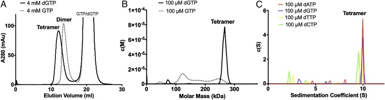
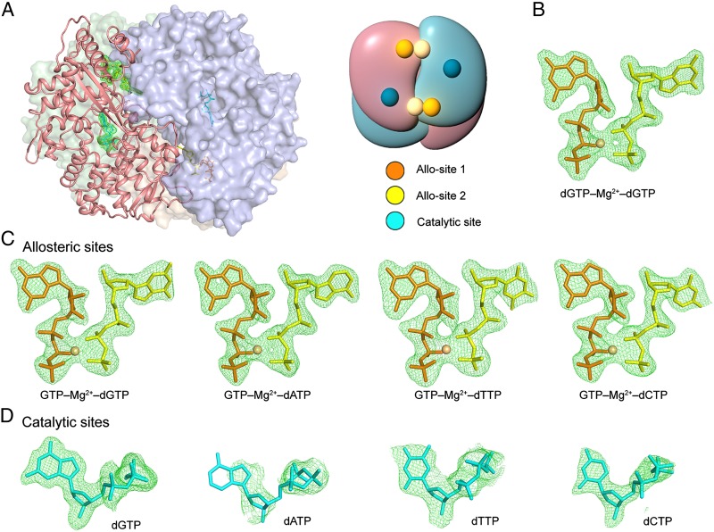
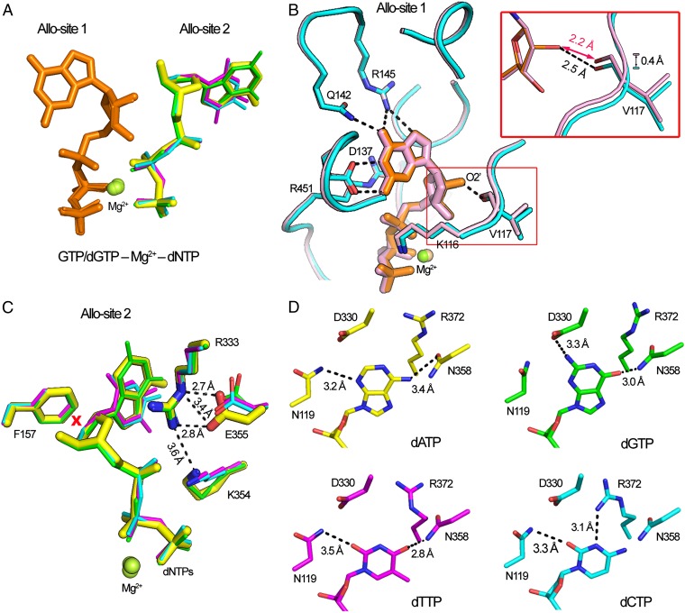
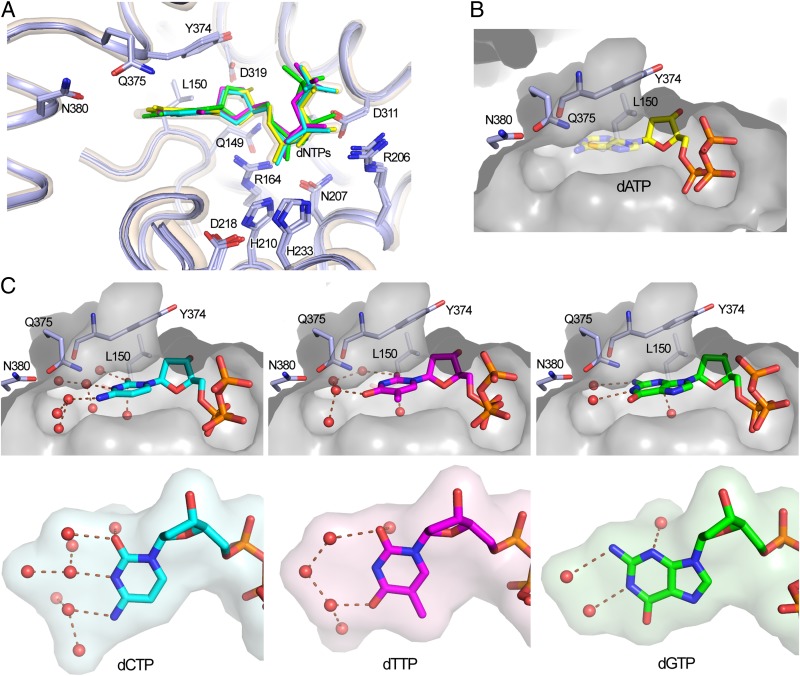
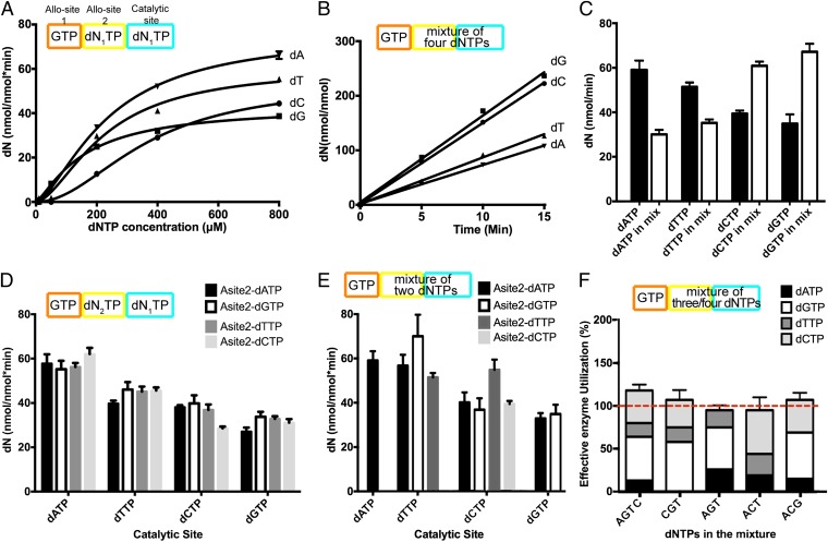
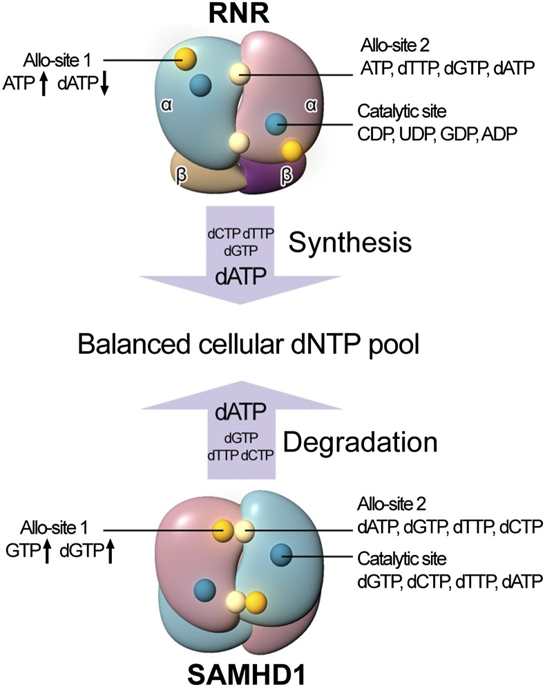
The sterile alpha motif and HD domain-containing protein 1 (SAMHD1), a dNTPase, prevents the infection of nondividing cells by retroviruses, including HIV, by depleting the cellular dNTP pool available for viral reverse transcription. SAMHD1 is a major regulator of cellular dNTP levels in mammalian cells. Mutations in SAMHD1 are associated with chronic lymphocytic leukemia (CLL) and the autoimmune condition Aicardi Goutières syndrome (AGS). The dNTPase activity of SAMHD1 can be regulated by dGTP, with which SAMHD1 assembles into catalytically active tetramers. Here we present extensive biochemical and structural data that reveal an exquisite activation mechanism of SAMHD1 via combined action of both GTP and dNTPs. We obtained 26 crystal structures of SAMHD1 in complex with different combinations of GTP and dNTP mixtures, which depict the full spectrum of GTP/dNTP binding at the eight allosteric and four catalytic sites of the SAMHD1 tetramer. Our data demonstrate how SAMHD1 is activated by binding of GTP or dGTP at allosteric site 1 and a dNTP of any type at allosteric site 2. Our enzymatic assays further reveal a robust regulatory mechanism of SAMHD1 activity, which bares resemblance to that of the ribonuclease reductase responsible for cellular dNTP production. These results establish a complete framework for a mechanistic understanding of the important functions of SAMHD1 in the regulation of cellular dNTP levels, as well as in HIV restriction and the pathogenesis of CLL and AGS.

Keywords: HIV restriction factor; allosteric regulation; dNTP metabolism; tetramerization; triphosphohydrolase.


Conflict of interest statement

The authors declare no conflict of interest.

Figures

Fig. 1.
Fig. 1.
GTP together with any dNTP, but not GTP alone, efficiently induces tetramerization of SAMHD1. (A) Size exclusion chromatograms of SAMHD1c-RN incubated with dGTP or GTP. Purified samples of SAMHD1c-RN (2 mg/mL, 200 μL) mixed with a final concentration of 4 mM dGTP or 4 mM GTP were applied to a Superdex 200 10/300 GL column. (B) Sedimentation velocity AUC results for SAMHD1c (1 mg/mL) with GTP (100 μM) or dGTP (100 μM). Little tetramer was formed with GTP alone. (C) Sedimentation velocity AUC results for SAMHD1c (1 mg/mL) with GTP (100 μM) and dNTP (100 μM) of any type.
Fig. 2.
Fig. 2.
Crystal structures of SAMHD1 tetramer in complex with GTP and a dNTP of any type. (A) Structure of SAMHD1 tetramer bound to nucleotides (Left). The four subunits are shown as ribbon or surface presentation, colored in light blue, pale cyan, salmon, and wheat, respectively. Nucleotides are shown as sticks. The representative allosteric and catalytic nucleotides in one subunit (salmon) were highlighted with green meshes. (Right) The schematic of the tetramer. (B–D) Unbiased Fo-Fc difference electron density (σ = 3.0) of the nucleotides bound to the SAMHD1 tetramer. (B) The allosteric site dGTP-Mg2+-dGTP (PDB ID code 4BZB). (C) The four scenarios of allosteric site GTP-Mg2+-dNTPs. (D) The dNTP substrates at the catalytic site.
Fig. 3.
Fig. 3.
The structures of the GTP-Mg2+-dNTPs at the allosteric sites. (A) Overlay of the structures of dGTP-Mg2+-dGTP and four dGTP-Mg2+-dNTPs. Nucleotides are shown as sticks and Mg2+ as spheres. Allo-site 1 nucleotides are colored in orange. Allo-site 2 nucleotides are colored individually according to dNTP types: dATP in yellow, dGTP in green, dTTP in magenta, and dCTP in cyan. (B) Superposition of Allo-site 1 structures of dGTP- or GTP-bound SAMHD1. SAMHD1 structures (pink: dGTP-bound, cyan: GTP-bound) are shown as ribbon with side chains of labeled residues shown as sticks. GTP and dGTP are shown in orange and pink, respectively. (Inset) Extra hydrogen bond (dashed line) between the 2’-hydroxyl group of GTP and main chain oxygen of V117. (C) Superposition of the structures of the four different dNTPs at Allo-site 2 of SAMHD1. dNTPs and labeled protein side chains are shown as sticks. The dATP-bound structure (yellow) is shown thicker than those of the other three. R333 stabilizes the bound dNTP through stacking interaction. The rigid side chain of F157 would cause steric clash (marked by a red X) with the 2′-OH group of an NTP. (D) The side chains of the surrounding residues adopt different conformations to adapt to the four different dNTP bases bound at Allo-site 2.
Fig. 4.
Fig. 4.
The structures of the dNTP substrates at the catalytic site. (A) Superposition of the structures of different dNTPs bound to the catalytic site of SAMHD1. The color scheme of the dNTPs is the same as in Fig. 3. The structure of the SAMHD1-dGTP complex is in semitransparent ribbon presentation (wheat). dNTPs and critical side chains were shown as sticks. (B) dATP binds weakly to the substrate-binding pocket (surface) with no clear conformation observed for the adenine base (semitransparent sticks). (C) The substrate-binding pockets (surface) and the bound dCTP, dTTP, and dGTP (sticks). Extensive water molecules (red spheres) at the dNTP base edges bridge the nonspecific interactions with the substrate-binding pocket. Perpendicular views of the base and water surfaces are shown in the lower panels. The pyrimidine bases (dCTP and dTTP) appear to have more extensive water networks than that of the purine base of dGTP.
Fig. 5.
Fig. 5.
Steady state kinetics for SAMHD1 dNTPase activity. All experiments were performed in triplicate. Schematic of the experimental setup for each assay is shown above the plots. (A) Single-dNTP substrate dNTPase assays. The dNTPase activity of SAMHD1c (0.5 μM) was assayed with indicated concentrations of dNTP, together with the same concentrations of GTP, for 5–15 min. The amount of dN product generated in the reactions was quantified by HPLC. (B) dNTPase assays with a mixture of all four dNTP substrates. Experiments were performed with a mixture of four dNTPs (500 μM each), together with SAMHD1c (0.5 μM) and GTP (500 μM). The linear initial rates of dN production were plotted. (C) Comparison of the dNTP hydrolysis rates in the single-substrate (black columns) and the four-substrate (white columns) assays at the dNTP(s) concentration of 500 μM. Each rate was normalized to that at the same concentration ratio of enzyme to total dNTPs. (D) dNTPase assays with preassembled SAMHD1 tetramers. SAMHD1c tetramers were preassembled by incubation with GTP and a particular Allo-site 2 dN2TP as indicated. After 100-fold dilution with reaction buffers containing a specific dN1TP substrate, the product was quantified and plotted as indicated. (E) dNTPase assays with pair-wise dNTP substrates. SAMHD1c (0.5 μM) activity assay were performed with GTP (500 μM) and a pair of dNTPs (500 μM each). The production rates of dNs were quantified and kappdN1-dN2 values were calculated as described in Materials and Methods. The rates are displayed as indicated. (F) Validation of the calculated kappdN2-dN1 values by the predicted effective enzyme utilization in the independent dNTPase assays with 3-dNTP or 4-dNTP mixtures. The kappdN2-dN1 values obtained in E lead to a calculated total effective enzyme utilization of 100% in each experiment, indicating the rates were correctly obtained.
Fig. 6.
Fig. 6.
SAMHD1 and RNR regulate cellular dNTP pool together. RNR and SAMHD1 are responsible for the production and degradation of dNTPs, respectively. Both of them have three specific nucleotides binding sites as indicated, with the types of the nucleotides that can occupy the sites marked on the side. The concentrations of cellular dNTPs are sensed and regulated by both of the enzymes. dATP is produced last by RNR and degraded last by SAMHD1.

References

    1. Goldstone DC, et al. HIV-1 restriction factor SAMHD1 is a deoxynucleoside triphosphate triphosphohydrolase. Nature. 2011;480(7377):379–382. - PubMed
    1. White TE, et al. The retroviral restriction ability of SAMHD1, but not its deoxynucleotide triphosphohydrolase activity, is regulated by phosphorylation. Cell Host Microbe. 2013;13(4):441–451. - PMC - PubMed
    1. Descours B, et al. SAMHD1 restricts HIV-1 reverse transcription in quiescent CD4(+) T-cells. Retrovirology. 2012;9:87. - PMC - PubMed
    1. Baldauf HM, et al. SAMHD1 restricts HIV-1 infection in resting CD4(+) T cells. Nat Med. 2012;18(11):1682–1687. - PMC - PubMed
    1. Laguette N, et al. SAMHD1 is the dendritic- and myeloid-cell-specific HIV-1 restriction factor counteracted by Vpx. Nature. 2011;474(7353):654–657. - PMC - PubMed

Publication types

LinkOut - more resources