. 2014 Oct 14;111(41):14924-9.
doi: 10.1073/pnas.1413706111. Epub 2014 Sep 29.

The hereditary spastic paraplegia-related enzyme DDHD2 is a principal brain triglyceride lipase

Affiliations

The hereditary spastic paraplegia-related enzyme DDHD2 is a principal brain triglyceride lipase

Jordon M Inloes et al. Proc Natl Acad Sci U S A. .

Abstract

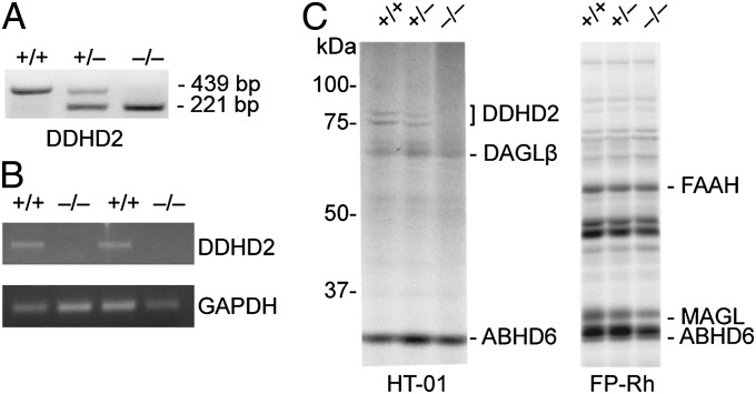
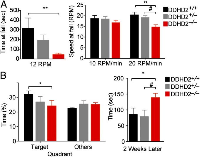
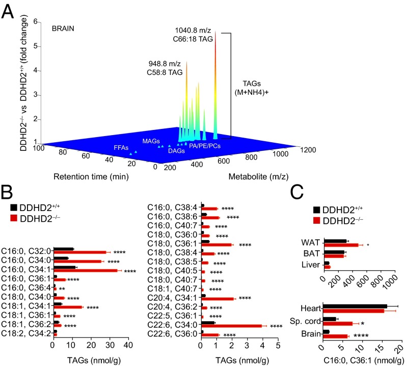
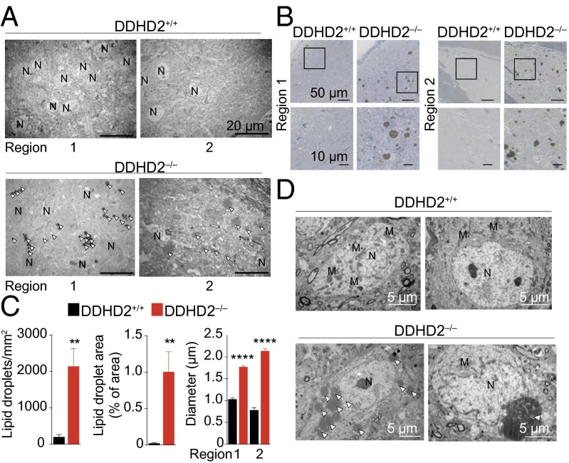
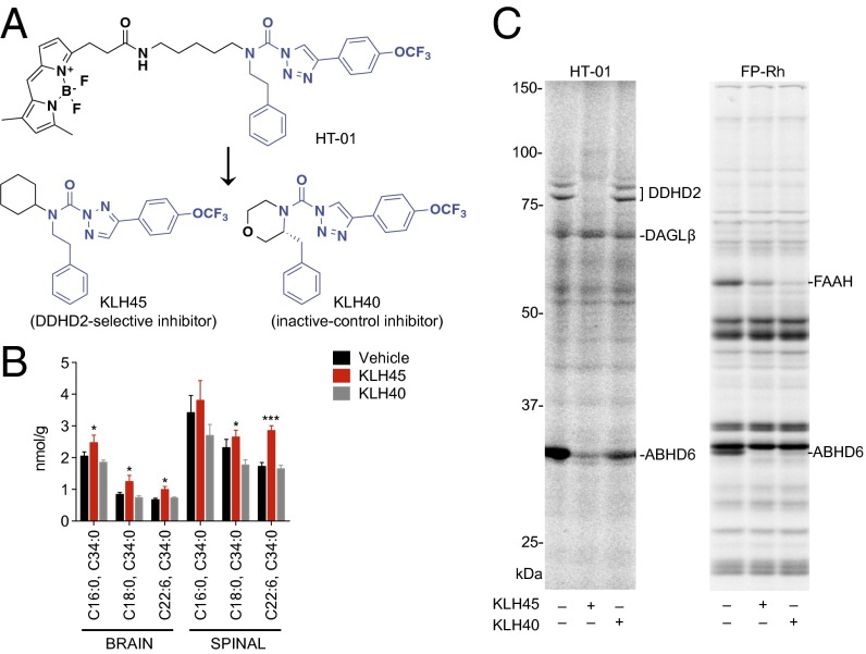
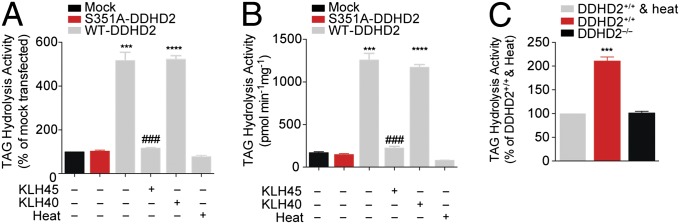
Complex hereditary spastic paraplegia (HSP) is a genetic disorder that causes lower limb spasticity and weakness and intellectual disability. Deleterious mutations in the poorly characterized serine hydrolase DDHD2 are a causative basis for recessive complex HSP. DDHD2 exhibits phospholipase activity in vitro, but its endogenous substrates and biochemical functions remain unknown. Here, we report the development of DDHD2(-/-) mice and a selective, in vivo-active DDHD2 inhibitor and their use in combination with mass spectrometry-based lipidomics to discover that DDHD2 regulates brain triglycerides (triacylglycerols, or TAGs). DDHD2(-/-) mice show age-dependent TAG elevations in the central nervous system, but not in several peripheral tissues. Large lipid droplets accumulated in DDHD2(-/-) brains and were localized primarily to the intracellular compartments of neurons. These metabolic changes were accompanied by impairments in motor and cognitive function. Recombinant DDHD2 displays TAG hydrolase activity, and TAGs accumulated in the brains of wild-type mice treated subchronically with a selective DDHD2 inhibitor. These findings, taken together, indicate that the central nervous system possesses a specialized pathway for metabolizing TAGs, disruption of which leads to massive lipid accumulation in neurons and complex HSP syndrome.


Conflict of interest statement

Conflict of interest statement: The authors declare competing financial interests. B.F.C. is cofounder and advisor for a biotechnology company interested in developing inhibitors for serine hydrolases as therapeutic targets.

Figures

Fig. 1.
Fig. 1.
Confirmation of loss of DDHD2 expression in DDHD2−/− mice. (A) Confirmation by PCR genotyping of germ-line transfer for an ES cell clone with targeted replacement of exon 8 of the Ddhd2 gene with a neomycin (Neo) selection cassette (see SI Appendix, Fig. S1 for gene targeting information). (B) Absence of DDHD2 mRNA in DDHD2−/− mice was confirmed by RT-PCR analysis of brain tissue. (C) Gel-based ABPP with the HT-01 probe confirmed lack of DDHD2 activity in brain membrane proteomes from DDHD2−/− mice. ABPP with the broad-spectrum probe FP-Rh confirmed that other serine hydrolase activities were unchanged in DDHD2−/− brain proteomes. [Note that DDHD2 is too low in abundance for detection by gel-based ABPP with the FP-Rh probe, but other representative serine hydrolases are marked for reference (17).].
Fig. 2.
Fig. 2.
Characterization of locomotor and cognitive functions in DDHD2−/− mice. (A) In a constant speed rotarod test, DDHD2−/− mice ran for a shorter time than DDHD2+/+ mice before falling (Left). In a rapidly accelerating speed rotarod test (Right), DDHD2−/− mice fell at lower speeds than DDHD2+/+ and DDHD2+/− mice. (B) In the Barnes maze spatial learning test, DDHD2−/− mice spent a shorter proportion of time in the quadrant with an escape chamber during training compared with DDHD2+/+ mice (Left). When the same mice were retested 2 wk later, the DDHD2−/− mice took longer to find the escape chamber compared with DDHD2+/+ and DDHD2+/− mice (Right). Data represent average values ± SEM. n = 13 mice per group. *P < 0.05 and **P < 0.01 for DDHD2+/+ versus DDHD2−/− mice; #P < 0.05 for DDHD2−/− versus DDHD2+/− mice.
Fig. 3.
Fig. 3.
TAG accumulation in DDHD2−/− brain tissue. (A) Lipidomic profile of brain tissue from DDHD2+/+ and DDHD2−/− mice. Metabolites were extracted from tissue of adult mice using a chloroform/methanol mixture and analyzed by LC–MS in both positive and negative ion mode as described in Materials and Methods. The XCMS algorithm identified features that were significantly different between DDHD2+/+ and DDHD2−/− brains, including a set of lipids that accumulated in DDHD2−/− brains and corresponded to the [M+NH4]+ adduct of triglycerides (TAGs). Fragmentation analysis and LC migration times were used to confirm structural assignment of TAGs (SI Appendix, Figs. S4 and S5). Other major lipid classes, including DAGs, monoglycerides (MAGs), free fatty acids (FFAs), and phospholipids [phosphatidic acids (PAs), phosphatidylcholines (PCs), and phosphatidylethanolamines (PEs)] were unchanged in DDHD2−/− brain tissue. (B) Targeted LC–MS analysis verified widespread elevations in TAGs in brain tissue from DDHD2−/− mice. (C) Comparison of TAGs in different tissues from DDHD2+/+ and DDHD2−/− mice (Sp. cord, spinal cord). Data represent average values ± SEM. n = 5–9 mice per group. *P < 0.05, **P < 0.01, ***P < 0.001, and ****P < 0.0001 for DDHD2−/− versus DDHD2+/+ mice.
Fig. 4.
Fig. 4.
Ultrastructural analysis of LD accumulation in DDHD2−/− brains. (A) Brains from adult DDHD2+/+ and DDHD2−/− mice were fixed, sectioned sagittally, and treated for 3 h with OsO4 before imaging with electron and light microscopy. Lower magnification images of thin (70 nm) sections of the pontine central gray (region 1) and septal nuclei (region 2) of 3-mo-old mice were captured by transmission EM, which revealed dark staining LDs in both regions in DDHD2−/− brains, but not in DDHD2+/+ brains. (Scale bar, 20 μm in all images.) (B) Thick, resin-embedded sections (2 μm) of the same regions were taken from 6- to 9-mo-old mice and counterstained with toluidine blue. Extensive LD accumulation was detected in both regions of DDHD2−/−, but not in DDHD2+/+ brains. (Scale bar, 50 μm in the Upper images and 10 μm in the Lower insets taken from the boxed regions of Upper images.) (C) The LD margins were identified using Image-Pro Plus 7.0 as described in Materials and Methods and used to quantify an increased number (Left), area (Center), and diameter (Right) of LDs in DDHD2−/− brains. (D) Ultrastructural EM analysis showing LD accumulation in neurons from DDHD2−/− mice. LD accumulation was observed either as multiple smaller droplets (Left) or one large droplet (Right) in the cytoplasm of neurons. Arrowhead denotes LDs. M, mitochondria; N, nucleus. See SI Appendix, Fig. S9 for additional images. Data represent average values ± SEM. n = 4 mice per group. **P < 0.01 and ****P < 0.0001 for DDHD2−/− versus DDHD2+/+ mice.
Fig. 5.
Fig. 5.
Mice treated subchronically with a selective DDHD2 inhibitor show TAG accumulation in the CNS. (A) Structural modifications to HT-01 yielded a DDHD2-selective inhibitor KLH45 and inactive-control inhibitor KLH40. (B) Targeted LC–MS analysis revealed accumulation of TAGs in brain and spinal cord (spinal) tissues from mice treated subchronically for 4 d with KLH45 versus vehicle or KLH40 (inhibitors were administered at 20 mg⋅kg−1 compound, i.p., every 12 h). (C) Competitive ABPP experiments confirmed the inactivation of DDHD2 in KLH45-treated but not KLH40-treated mice. Both KLH45 and KLH40 partially inhibited ABHD6 and FAAH but showed negligible cross-reactivity with other brain serine hydrolases. Data represent average values ± SEM. n = 4 mice per group. *P < 0.05 and ***P < 0.001 for KLH45-treated versus vehicle-treated mice.
Fig. 6.
Fig. 6.
DDHD2 exhibits TAG hydrolase activity. (A and B) Soluble lysates from HEK293T cells transiently transfected with a WT–DDHD2 cDNA showed greater C18:1/C18:1/C18:1 TAG hydrolytic activity measured by either a radiolabeled TLC (A) or LC–MS (B) assay compared with lysates from mock-transfected cells, heat-denatured WT–DDHD2-transfected lysates, or cells transfected with an S351A–DDHD2 mutant cDNA. Both assays report formation of C18:1 fatty acid. For measurement of 14C-C18:1 MAG and 14C-C18:1/C18:1 DAG formation in the radiolabeled 14C-TAG substrate assay, see SI Appendix, Fig. S15. In both substrate assays, KLH45 but not KLH40 blocked the TAG hydrolase activity of DDHD2. (C) Soluble brain lysates from DDHD2−/− mice show reduced TAG hydrolysis activity compared with soluble brain lysates from DDHD2+/+ mice measured by a radiolabeled substrate assay following conversion of C18:1/C18:1/C18:1 TAG to C18:1 fatty acid. Heat-denatured DDHD2+/+ brain lysates were assayed as a control and displayed a similar signal to those observed in DDHD2−/− lysates. Data represent average values ± SEM for three experimental replicates per group. ***P < 0.001 and ****P < 0.0001 for WT–DDHD2 versus S351A–DDHD2 transfected groups or DDHD2+/+ versus DDHD2−/− groups; ###P < 0.001 for KLH45-treated versus DMSO-treated WT–DDHD2 groups.

References

    1. Rabbani B, Mahdieh N, Hosomichi K, Nakaoka H, Inoue I. Next-generation sequencing: Impact of exome sequencing in characterizing Mendelian disorders. J Hum Genet. 2012;57(10):621–632. - PubMed
    1. Fink JK. Hereditary spastic paraplegia: Clinico-pathologic features and emerging molecular mechanisms. Acta Neuropathol. 2013;126(3):307–328. - PMC - PubMed
    1. Novarino G, et al. Exome sequencing links corticospinal motor neuron disease to common neurodegenerative disorders. Science. 2014;343(6170):506–511. - PMC - PubMed
    1. Rainier S, et al. Neuropathy target esterase gene mutations cause motor neuron disease. Am J Hum Genet. 2008;82(3):780–785. - PMC - PubMed
    1. Tesson C, et al. Alteration of fatty-acid-metabolizing enzymes affects mitochondrial form and function in hereditary spastic paraplegia. Am J Hum Genet. 2012;91(6):1051–1064. - PMC - PubMed

Publication types

MeSH terms