. 2014 Oct 14;111(41):E4350-8.
doi: 10.1073/pnas.1417138111. Epub 2014 Sep 29.

Transmembrane domain of surface-exposed outer membrane lipoprotein RcsF is threaded through the lumen of β-barrel proteins

Affiliations

Transmembrane domain of surface-exposed outer membrane lipoprotein RcsF is threaded through the lumen of β-barrel proteins

Anna Konovalova et al. Proc Natl Acad Sci U S A. .

Abstract

RcsF (regulator of capsule synthesis) is an outer membrane (OM) lipoprotein that functions to sense defects such as changes in LPS. However, LPS is found in the outer leaflet, and RcsF was thought to be tethered to the inner leaflet by its lipidated N terminus, raising the question of how it monitors LPS. We show that RcsF has a transmembrane topology with the lipidated N terminus on the cell surface and the C-terminal signaling domain in the periplasm. Strikingly, the short, unstructured, charged transmembrane domain is threaded through the lumen of β-barrel OM proteins where it is protected from the hydrophobic membrane interior. We present evidence that these unusual complexes, which contain one protein inside another, are formed by the Bam complex that assembles all β-barrel proteins in the OM. The ability of the Bam complex to expose lipoproteins at the cell surface underscores the mechanistic versatility of the β-barrel assembly machine.

Keywords: envelope stress response; membrane biogenesis; protein folding; signal transduction.


Conflict of interest statement

The authors declare no conflict of interest.

Figures

Fig. 1.
Fig. 1.
RcsF is a surface-exposed lipoprotein. (A) RcsF is detected on the cell surface in a dot blot assay. Whole cells or cell lysates of a WT or rcsF strain were spotted on the nitrocellulose membrane and probed with corresponding antibodies. For OM-GFP, the expression plasmid was introduced into the indicated strains. (B) Surface exposure of RcsF depends on OM targeting signal sequence. Whole cells expressing the indicated RcsF variants were probed with anti-RcsF antibodies in a dot blot assay (Upper) and total RcsF protein levels were analyzed by an immunoblot assay with anti-RcsF antibodies. (C) The N terminus of RcsF is surface exposed. RcsFWT and RcsF-FLAG variants were expressed from a plasmid and whole cells or cell lysates were probed with anti-FLAG, anti-RcsF and anti-BamC (as a control of cell envelope integrity) antibodies in a dot blot assay. Total RcsF protein levels were analyzed by an immunoblot assay with anti-RcsF antibodies. RcsF-FLAG variants are indicated by the amino acid number after which the FLAG tag was introduced.
Fig. 2.
Fig. 2.
RcsF interacts with OMPs and BamA in a distinct fashion. (A) Site-specific cross-linking of RcsF to OmpA, OmpC, OmpF, and BamA. RcsF-Strep pBPA variants and RcsF-Strep (negative control) were expressed from a plasmid and cells were subjected to UV cross-linking. Presence of cross-linked product was analyzed by a size shift of the protein of interest with corresponding antibodies. pBPA variants are named by the amino acid number at which pBPA was introduced. Note, OmpC and OmpF are homologs and are both recognized by anti-OmpF antibodies and cannot be resolved on the standard SDS/PAGE gel. Anti-OmpA antibodies also cross-react with LamB. (B) Sites of interaction of RcsF with OMPs and BamA on the RcsF structure [Protein Data Bank (PDB) ID code 2L8Y]. Sites of cross-linking to OMPs are shown in red, and sites of cross-linking to BamA are shown in green. Note the RcsF linker is truncated in this structure and the molecule starts at amino acid P34. (C) Sites of interaction of RcsF with OMPs and BamA on the linear RcsF sequence and corresponding secondary structure elements. Sites are color coded as in B.
Fig. 3.
Fig. 3.
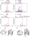
RcsF G60pBPA cross-links inside the lumen of OmpC and OmpF. Culture expressing RcsF-Strep G60pBPA was subjected to in vivo UV cross-linking. RcsF-Strep G60pBPA complexes were affinity purified and resolved on SDS/PAGE. Cross-linked peptides were rendered by chymotryptic digestion and subjected to high-resolution MS-based proteomic analysis. A and B contain MS/MS spectra assigned to RcsF-OmpC and RcsF-OmpF cross-linked peptide species, with their b-and y-type fragment ions indicated in red and blue, respectively. Prominent ions are labeled with their empirical m/z values and b or y ion assignments. All matched fragment ions are additionally represented as flags on the sequence diagrams above the spectra. The pBPA residue is indicated by X. (C) Periplasmic and side view of the extracellular loop 3 in the lumen of the OmpC and OmpF barrel, based on the crystal structures (PDB ID codes 2J1N and 2ZFG, respectively). Sites of RcsF cross-linking identified in A and B are highlighted in red.
Fig. 4.
Fig. 4.
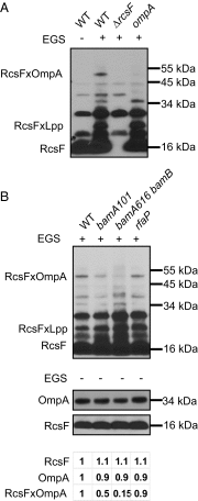
Mutations in the Bam complex affect levels of RcsF/OmpA complexes in vivo. (A) Cells of indicated strains were subjected to in vivo cross-linking with EGS and analyzed by immunoblot with anti-RcsF antibodies. The cross-linked RcsFxOmpA band has the expected size of 50 kDa. (B) In vivo cross-linking with EGS (Upper) performed as in A. Total OmpA and RcsF levels in each strain were analyzed in un–cross-linked samples (Lower) by anti-OmpA and anti-RcsF antibodies. Intensity of the OmpA, RcsF, and RcsF/OmpA bands were quantified by ImageJ and normalized to the WT.
Fig. 5.
Fig. 5.
RcsF/OmpA complexes are formed during OmpA folding in vitro. (A) Urea denatured FLAG-OmpA was refolded in TBS with 0.1% Triton X-100 for 2 h and then an equimolar amount of purified RcsF-Strep K40pBPA (positive for in vivo cross-linking), and RcsF-Strep D72pBPA (negative for in vivo cross-linking) was added; an equal volume of buffer was added to a control reaction (−). Reactions were incubated for additional 2 h and then subjected to UV cross-linking. Reactions were stopped by the addition of SDS-loading buffer and split into unboiled (−) and boiled (+) samples and analyzed by immunoblot for the presence of the RcsFxOmpA band with anti-FLAG antibodies. Note, folded OmpA (f) remains folded in −boil samples and migrates faster on SDS/PAGE than unfolded OmpA (u). (B) Urea denatured FLAG-OmpA was refolded in TBS with 0.1% Triton X-100 in the absence (−) or presence of an equimolar amount of the corresponding RcsF-Strep pBPA variant for 2 h. Samples were UV cross-linked and treated as described in A.
Fig. 6.
Fig. 6.
Proposed model of the mechanism of surface localization of RcsF. RcsF adopts Outside-In orientation traversing OM via the β-barrel lumen of an OMP. This conformation requires formation of the RcsF/OMP complexes during OMP folding by Bam machine.

References

    1. Silhavy TJ, Kahne D, Walker S. The bacterial cell envelope. Cold Spring Harb Perspect Biol. 2010;2(5):a000414. - PMC - PubMed
    1. Nikaido H. Molecular basis of bacterial outer membrane permeability revisited. Microbiol Mol Biol Rev. 2003;67(4):593–656. - PMC - PubMed
    1. Ricci DP, Silhavy TJ. The Bam machine: A molecular cooper. Biochim Biophys Acta. 2012;1818(4):1067–1084. - PMC - PubMed
    1. Tokuda H, Matsuyama S. Sorting of lipoproteins to the outer membrane in E. coli. Biochim Biophys Acta. 2004;1694(1-3):IN1–IN9. - PubMed
    1. Hayashi S, Wu HC. Lipoproteins in bacteria. J Bioenerg Biomembr. 1990;22(3):451–471. - PubMed

Publication types

MeSH terms

LinkOut - more resources