. 2014 Nov 4;111(44):15827-32.
doi: 10.1073/pnas.1416751111. Epub 2014 Sep 29.

Marine algae and land plants share conserved phytochrome signaling systems

Affiliations

Marine algae and land plants share conserved phytochrome signaling systems

Deqiang Duanmu et al. Proc Natl Acad Sci U S A. .

Abstract

Phytochrome photosensors control a vast gene network in streptophyte plants, acting as master regulators of diverse growth and developmental processes throughout the life cycle. In contrast with their absence in known chlorophyte algal genomes and most sequenced prasinophyte algal genomes, a phytochrome is found in Micromonas pusilla, a widely distributed marine picoprasinophyte (<2 µm cell diameter). Together with phytochromes identified from other prasinophyte lineages, we establish that prasinophyte and streptophyte phytochromes share core light-input and signaling-output domain architectures except for the loss of C-terminal response regulator receiver domains in the streptophyte phytochrome lineage. Phylogenetic reconstructions robustly support the presence of phytochrome in the common progenitor of green algae and land plants. These analyses reveal a monophyletic clade containing streptophyte, prasinophyte, cryptophyte, and glaucophyte phytochromes implying an origin in the eukaryotic ancestor of the Archaeplastida. Transcriptomic measurements reveal diurnal regulation of phytochrome and bilin chromophore biosynthetic genes in Micromonas. Expression of these genes precedes both light-mediated phytochrome redistribution from the cytoplasm to the nucleus and increased expression of photosynthesis-associated genes. Prasinophyte phytochromes perceive wavelengths of light transmitted farther through seawater than the red/far-red light sensed by land plant phytochromes. Prasinophyte phytochromes also retain light-regulated histidine kinase activity lost in the streptophyte phytochrome lineage. Our studies demonstrate that light-mediated nuclear translocation of phytochrome predates the emergence of land plants and likely represents a widespread signaling mechanism in unicellular algae.

Keywords: light harvesting; light signaling evolution; marine ecology; phytoplankton; transcriptomics.


Conflict of interest statement

The authors declare no conflict of interest.

Figures

Fig. 1.
Fig. 1.
Domain structures of phytochrome proteins. The N-terminal photosensory core module (PCM) of phytochromes is composed of PAS, GAF, and PHY domains (dashed box). Colors on the chromophore binding GAF domains correspond to those of the two reversibly photointerconverting states of each phytochrome where known or as described here. C-terminal output modules of phytochromes from all Archaeplastida lineages typically contain one or two PAS domains adjacent to histidine kinase modules (HKM). Lack of C-terminal receiver (REC) domains in streptophyte phytochromes contrasts with their presence in prasinophyte, glaucophyte, and cryptophyte phytochromes. Structurally distinct phytochrome eukaryotic kinase (PEK) hybrids are present in the cryptophyte alga Guillardia theta. The Ectocarpus siliculosus photocycle shown here (asterisk) may not be representative of other heterokont phytochrome photocycles. Domain names: CHD, cyclase homology domain; GAF, cGMP phosphodiesterase/adenylate cyclase/FhlA; H/KD, HisKA and H-ATPase-c domains comprising the HKM; PAS, Per/Arnt/Sim; PHY, phytochrome; PKC, protein kinase catalytic domain; REC, response regulator receiver; and RING, really interesting new gene. Taxonomic assignments (colored bars) follow color-coding used in Fig. 2. Dashed outlines indicate domains that are not always present.
Fig. 2.
Fig. 2.
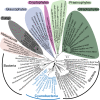
Evolutionary analyses establish common ancestry of phytochromes from Archaeplastida (and cryptophyte) lineages and support presence in early eukaryotes. Evolutionary relationships are based on maximum likelihood (ML) analyses of phytochromes from 128 representative taxa using 407 homologous positions in the N-terminal PAS–GAF–PHY region. Colored backgrounds indicate eukaryotic sequences. Cyanobacterial sequences are in blue text. Collapsed streptophyte clades are named according to Arabidopsis thaliana (if present) but also include other taxa (SI Appendix, Fig. S3). Plastids in cryptophyte and heterokont algae were likely attained through independent secondary endosymbiosis with red algae (23), unlike Archaeplastidal plastids, which are thought to have arisen from a single primary endosymbiosis event with cyanobacteria (8, 9). Placement of cryptophyte PCMs within the Archaeplastida could therefore represent a red algal version recruited via EGT from the secondary plastid ancestor (although absent from sequenced red algal genomes) or contributions from a putative green algal forebear implicated in its genome composition (8). The tree is unrooted. Support is indicated by open circles (≥90%, ML; ≥0.9 posterior probability, Bayesian) or black circles (≥75%, ML; ≥0.9 posterior probability, Bayesian).
Fig. 3.
Fig. 3.
Synchronized M. pusilla cells exhibit strong predawn phytochrome gene expression, preceding most photosynthesis genes and phytochrome protein translocation to nucleus. (A) Micromonas cells in mid-exponential growth exhibit synchronized division once per day. Cell size (green circles, represented as bead-normalized mean forward angle light scatter, FALS) increases throughout the photoperiod as cells prepare to divide and decreases (green arrow) once division begins at the onset of night (black arrow). Total cell divisions (bars) are shown since the start of the experiment. Division progresses into predawn hours and a second round commences at the end of the day 2 photoperiod (second black arrow). Immunoblot quantitation of MpPHY protein shows little variation from the first measurement (0.5 h before lights on), as determined from biological triplicates and normalized against alpha-tubulin (reported as fold change ± SD). (B) Immunoblot analysis of total (T) and nucleus-localized (N) MpPHY during the light period (T1P and T2P), the subsequent dark period (T3P), and the following morning (T4P). Numbers over lanes indicate nucleus-localized MpPHY protein fold changes relative to T4P, the earliest light period time point (as done in A) and normalized against RNA polymerase II (RNAP II). Bars and error bars represent the mean and SD of technical duplicates, respectively. (C) MpPHY and MpPUBS transcript abundances over the diel. Bars represent average quartile normalized fragments per kilobase of transcript per million mapped reads (FPKM) from biological triplicates and error bars represent the SD. “R” in the sample name indicates RNA time points. T tests between adjacent time points show significance (*, ** represent significance for comparisons with the preceding time point; symbols over T1R data represent a test between T4R, just as lights came on, and T1R). (D) Z-score analysis of MpPHY, heme oxygenases (HMOX1 and HMOX2, responsible for initial chromophore synthesis steps), FDBRs, and photosynthesis-related genes (the latter in nonbold font). Relative change from mean transcript levels (log transformed) in negative (blue) or positive (red) directions is shown for each gene across time points. Upper-quartile normalized FPKM (± SD) are provided in SI Appendix, Table S4.
Fig. 4.
Fig. 4.
Spectral properties and light-regulated protein kinase activities of recombinant prasinophyte phytochrome. (A) Spectral properties of the PCM of M. pusilla phytochrome (MpPHY-PCM). (B) Spectra of D. tenuilepis (DtPHY-PCM) and A. thaliana phytochrome A (AtPHYA-PCM) PCMs. Presence of a histidine kinase domain following the PCM (DtPHY-ΔL, DtPHY-ΔL H927Q) does not change D. tenuilepis phytochrome spectral properties (the three spectra overlap). The ∆L truncation of DtPHY lacks all three REC and CHD domains (Fig. 1), as does DtPHY-ΔL H927Q, which also lacks the conserved histidine autophosphorylation site. (C) Comparative kinase analysis of dark-adapted states of DtPHY-ΔL (Po), DtPHY-ΔL with single mutation (H927Q), and Synechocystis Cph1 (Pr) and their respective far-red absorbing Pfr states. Top, Middle, and Bottom represent autoradiograph, zinc blot, and Coomassie blue-stained images of the transblotted proteins (5 μg per lane). Numbers indicate kinase activity relative to Cph1 Pr by normalization of each sample against the zinc blot signal. Technical duplicates were analyzed.

Comment in

References

    1. Chen M, Chory J, Fankhauser C. Light signal transduction in higher plants. Annu Rev Genet. 2004;38:87–117. - PubMed
    1. Giraud E, Verméglio A. Bacteriophytochromes in anoxygenic photosynthetic bacteria. Photosynth Res. 2008;97(2):141–153. - PubMed
    1. Rodriguez-Romero J, Hedtke M, Kastner C, Müller S, Fischer R. Fungi, hidden in soil or up in the air: Light makes a difference. Annu Rev Microbiol. 2010;64:585–610. - PubMed
    1. Auldridge ME, Forest KT. Bacterial phytochromes: More than meets the light. Crit Rev Biochem Mol Biol. 2011;46(1):67–88. - PubMed
    1. Mathews S. Phytochrome-mediated development in land plants: Red light sensing evolves to meet the challenges of changing light environments. Mol Ecol. 2006;15(12):3483–3503. - PubMed

Publication types

MeSH terms