. 2014 Dec;21(6):673-83.
doi: 10.1093/dnares/dsu029. Epub 2014 Sep 28.

The genome landscape of the african green monkey kidney-derived vero cell line

Affiliations

The genome landscape of the african green monkey kidney-derived vero cell line

Naoki Osada et al. DNA Res. 2014 Dec.

Abstract

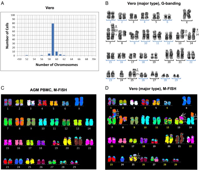
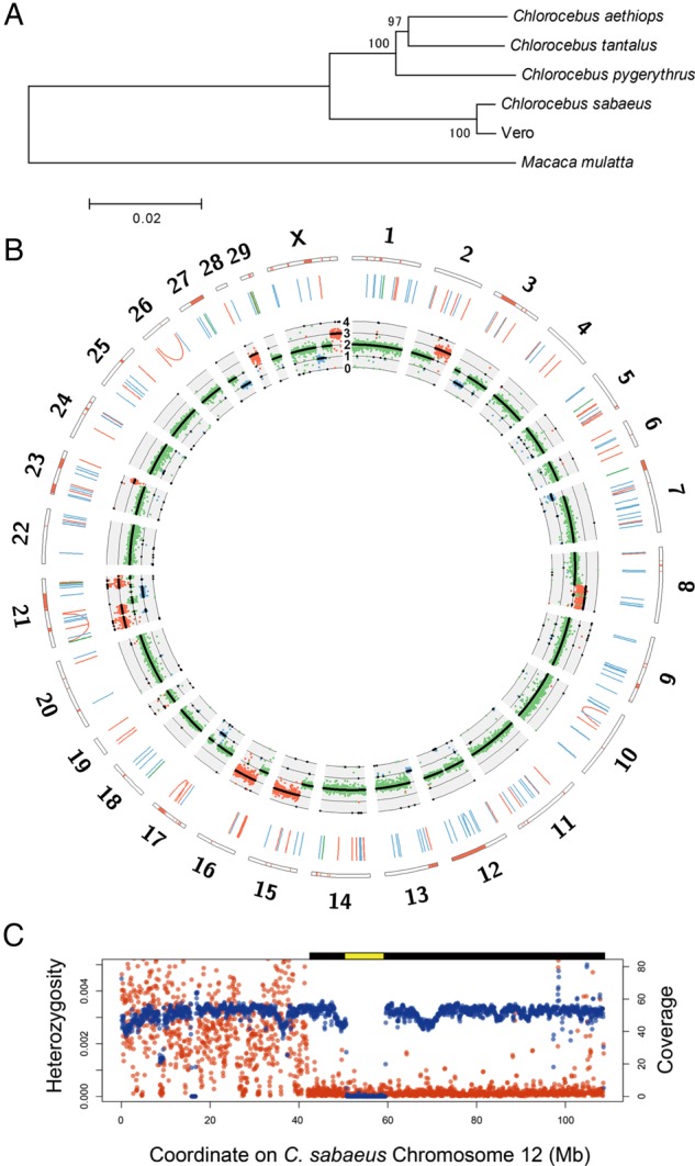
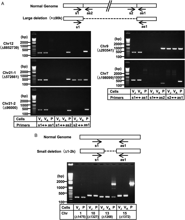
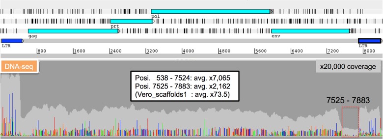
Continuous cell lines that originate from mammalian tissues serve as not only invaluable tools for life sciences, but also important animal cell substrates for the production of various types of biological pharmaceuticals. Vero cells are susceptible to various types of microbes and toxins and have widely contributed to not only microbiology, but also the production of vaccines for human use. We here showed the genome landscape of a Vero cell line, in which 25,877 putative protein-coding genes were identified in the 2.97-Gb genome sequence. A homozygous ∼9-Mb deletion on chromosome 12 caused the loss of the type I interferon gene cluster and cyclin-dependent kinase inhibitor genes in Vero cells. In addition, an ∼59-Mb loss of heterozygosity around this deleted region suggested that the homozygosity of the deletion was established by a large-scale conversion. Moreover, a genomic analysis of Vero cells revealed a female Chlorocebus sabaeus origin and proviral variations of the endogenous simian type D retrovirus. These results revealed the genomic basis for the non-tumourigenic permanent Vero cell lineage susceptible to various pathogens and will be useful for generating new sub-lines and developing new tools in the quality control of Vero cells.

Keywords: Vero cell; animal cell substrate; infectious diseases; vaccine; whole genome.


Figures

Figure 1.
Figure 1.
Karyotyping of the Vero JCRB0111 cell line. (A) Chromosome number in the Vero cell line based on 100 Giemsa-stained metaphases, which showed that the modal number was 59 chromosomes and differences in the chromosome number between 52 and 62 indicated heterogeneous karyotypes. (B) G-banded karyotype of Vero cells with 59 chromosomes consisted of 16 homologous pairs (blue numbers) and 13 abnormal chromosomes (black numbers). (C and D) M-FISH signal pattern using human probes on a normal AGM metaphase (C), and a main clone of the Vero cell line (D). Paints of human chromosomes 1–22 and X showed homologous regions in the AGF and the identity of 32 and 40 syntenic blocks in a normal female and Vero, respectively. The number on the right of aberrant chromosomes (D) shows correspondence to AGF chromosomes. The derivative chromosome der(13) (white arrow in D) showed an extra signal that corresponded to AGM chromosomes 15 and 22, both of which were painted by human chromosome 3. Although the addition in der(13) could not be distinguished using human M-FISH probes, the copy number analysis revealed a gain at chromosome 15q (Fig. 2B), which indicated that der(13) had additional material over chromosome 15.
Figure 2.
Figure 2.
Genome landscape of the Vero genome. (A) Phylogeny of mitochondrial genomes of the Vero cell line, four Chlorocebus species, and Macaca mulatta. Bootstrap values with 1,000 replications were shown upon the branches. (B) Circos plot of the Vero cell genome. The orange bars in the outermost rectangles represent LOH regions. The blue, green, and orange lines in the middle layer show deletions, duplications, and inversions larger than 1 kb, respectively. The innermost plot shows the coverage of paired-end reads and expected ploidy (black lines). The blue, green, and orange dots represent the coverage values in 1×, 2×, and 3× regions, respectively. (C) The large deletion and LOH regions on Chlorocebus sabaeus chromosome 12. The red and blue points represent average heterozygosity (the number of heterozygous SNVs per bp) and genome coverage of paired-end reads in 1-Mb-size windows, respectively. The predicted homozygous deletion regions and LOH regions are shown as yellow and black bars on the plot area, respectively.
Figure 3.
Figure 3.
Validation of genomic deletions in Vero cells by PCR analysis. Large deletions (>90 kb deletions) (A) and some small deletions (1–2 kb deletions) (B) were selected. Although the genomic PCR confirmed the existence of breakpoint junctions for the 573 kb deletion in chromosome 21 and the 294 kb deletion in chromosome 9, a part of these regions appeared to exist somewhere in the genome (A; see also Supplementary Fig. S2). Amplicons corresponding to the deletions predicted in chromosomes 1 and 10 were produced not only from Vero cells, but also from AGM PBMC, while amplicons corresponding to the ‘non-deleted’ counterparts were not produced even from AGM PBMC (B; see also Supplementary Fig. S2), which indicated that our determined sequences for the Vero cell genome existed homozygously in these regions not only in Vero cells, but also in AGM PBMC. This paradox might be attributed to the possible incompleteness of the currently available version of the AGM whole-genome draft sequence or polymorphic state of the deletion within AGM populations. Normal Genome indicates the sequences predicted from the draft genome sequences of AGM and the rhesus macaque. Arrows indicate the primer positions used in the PCR analyses. The ‘Δ’ indicates the genomic deletion size predicted by the massively parallel sequencing system. The templates used were as follows: VJ, Vero JCRB0111; VA, Vero ATCC; P, AGM PBMC. PCR amplicons were sequenced to confirm the breakpoint sequences, which are shown in Supplementary Fig. S2. Chr, chromosome.
Figure 4.
Figure 4.
Characterization of proviral SRV sequences. DNA-seq short read mapping to the complete SRV-Vero genome sequence. Read depth and mismatch nucleotides are shown in the following colours (Depth: light grey, A: light green, T: red, G: orange, C: dark blue). The high variability of SRV sequences was rarely detected in the 7525–7883 nt region, whereas high variability was observed throughout other regions.

References

    1. Yasumura Y., Kawakita Y. Studies on SV40 in tissue culture: preliminary step for cancer reserach in vitro (in Japanese) Nihon Rinsho. 1963;21:1201–15.
    1. Yasumura Y., Kawakita Y. Studies on SV40 in tissue culture: preliminary step for cancer research in vitro. In: Simizu, B., Terasima T., editors. VERO cells: origin, properties and biomedical applications. Chiba, Japan: Department of Microbiology School of Medicine Chiba University; 1988. pp. 1–19.
    1. Sasaki K., Makino S., Kasahara S. Studies on measles virus. II. Propagation in two established simian renal cell lines and development of a plaque assay. Kitasato Arch. Exp. Med. 1964;37:27–42. - PubMed
    1. Rhim J.S., Schell K. Cytopathic and plaque assay of rubella virus in a line of African green monkey kidney cells (Vero) Proc. Soc. Exp. Biol. Med. 1967;125:602–6. - PubMed
    1. Liebhaber H., Riordan J.T., Horstmann D.M. Replication of rubella virus in a continuous line of African green monkey kidney cells (Vero) Proc. Soc. Exp. Biol. Med. 1967;125:636–43. - PubMed

Publication types