. 2014 Aug 1;9(4):478-498.
doi: 10.1016/j.nantod.2014.06.003.

Nanomedicine in the Management of Microbial Infection - Overview and Perspectives

Affiliations

Nanomedicine in the Management of Microbial Infection - Overview and Perspectives

Xi Zhu et al. Nano Today. .

Abstract

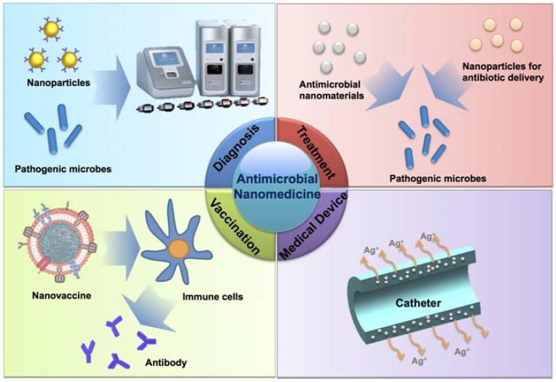
For more than 2 billion years, microbes have reigned on our planet, evolving or outlasting many obstacles they have encountered. In the 20th century, this trend took a dramatic turn with the introduction of antibiotics and vaccines. Nevertheless, since then, microbes have progressively eroded the effectiveness of previously successful antibiotics by developing resistance, and many infections have eluded conventional vaccine design approaches. Moreover, the emergence of resistant and more virulent strains of bacteria has outpaced the development of new antibiotics over the last few decades. These trends have had major economic and health impacts at all levels of the socioeconomic spectrum - we need breakthrough innovations that could effectively manage microbial infections and deliver solutions that stand the test of time. The application of nanotechnologies to medicine, or nanomedicine, which has already demonstrated its tremendous impact on the pharmaceutical and biotechnology industries, is rapidly becoming a major driving force behind ongoing changes in the antimicrobial field. Here we provide an overview on the current progress of nanomedicine in the management of microbial infection, including diagnosis, antimicrobial therapy, drug delivery, medical devices, and vaccines, as well as perspectives on the opportunities and challenges in antimicrobial nanomedicine.

Keywords: Diagnosis; Drug delivery; Medical device; Microbial infection; Nanomedicine; Therapy; Vaccine.


Figures

Figure 1
Figure 1
Nanomedicine applications in the management of microbial infection.
Figure 2
Figure 2
(A) Assay workflow for detection of Candida with T2MR. (B) Schematic depicting the T2MR detection particle reagent. Oligonucleotide probes are covalently conjugated to SPIONs. For each target, two populations of nanoparticles were generated, each bearing a distinct target-complementary probe. Upon hybridization to the target strand amplified in excess by asymmetric PCR, these nanoparticles cluster, leading to a change of the sample’s T2MR signal. The extent of clustering increases with the target DNA concentration. Reprinted with permission from [19]. Copyright 2013, American Association for the Advancement of Science.
Figure 3
Figure 3
Bio-barcode assay for DNA and protein detection. Schematic representation of (A) protein detection using the bio-barcode assay; (B) nucleic acid detection using the bio-barcode assay; and (C) the scanometric detection method. Au-NP, gold nanoparticle; MMP, magnetic microparticle. Reprinted with permission from[29]. Copyright 2006, Nature Publishing Group.
Figure 4
Figure 4
Schematic of (A) nanomaterials with inherent antimicrobial properties, and (B) nanoparticle-based antimicrobial drug delivery systems.
Figure 5
Figure 5
Chemical structure of (A) the designed peptide with cholesterol, glycine, arginine, and TAT; and (C) cationic amphiphilic polycarbonate. (B) and (D) are the formation of micelles of (A) and (C), respectively, simulated through molecular modeling using Materials Studio software. Reprinted with permission from [76] and [80]. Copyright 2009, Nature Publishing Group [76]. Copyright 2011, Nature Publishing Group [80].
Figure 6
Figure 6
(A) Schematic representation of the designed nanoparticle-mediated drug targeting to bacterial cell walls. The nanoparticles avoid uptake or binding to nontarget cells or blood components at physiologic pH 7.4 due to a slight negative charge and surface PEGylation. The weakly acidic conditions at sites of certain infections activate the surface charge-switching mechanism, resulting in nanoparticle binding to negatively charged bacteria. (B) Nanoparticle zeta potential vs. pH demonstrates notable switching from anionic to cationic with decreases in pH in PLGA-PLH-PEG but not PLGA-PEG nanoparticles. (C) Minimum inhibitory concentrations (MIC) of the different vancomycin formulations in S. aureus. Reprinted with permission from [120]. Copyright 2012, American Chemical Society.
Figure 7
Figure 7
Strategies of the application of antimicrobial nanomaterials in medical devices, including (A) surface coating by direct deposition, (B) surface coating by blending antibacterial nanomaterials into a polymer coating, and (C) embedding of antimicrobial nanomaterials into the bulk matrix of medical devices.
Figure 8
Figure 8
Mechanisms by which nanoparticles alter the induction of immune responses. Reprinted with permission from [158]. Copyright 2013, Nature Publishing Group.
Figure 9
Figure 9
(A) Schematic preparation of nanoparticle-detained toxins, consisting of substrate-supported RBC membranes into which PFTs can spontaneously incorporate.(B) TEM image of the particle vectors with uranyl-acetate staining (scale bar, 80 nm). (C) Live, whole-body fluorescent imaging of nanotoxoid(Hla) at 1h after subcutaneous administration. (D) Anti-Hla IgG titres at day 21 (n=7). Black lines indicate geometric means. Anti-Hla titres from mice vaccinated with non-toxin loaded particle vectors (nanotoxoid(-)) were monitored as controls (open triangles). (E) Unvaccinated mice (black triangles, solid line) and mice vaccinated with heat-treated Hla (prime; blue squares, dashed line), nanotoxoid(Hla) (prime; blue circles, solid line), heat-treated Hla (prime tboost; red squares, dashed line) or nanotoxoid(Hla) (prime + boost; red circle, solid line) received intravenous or subcutaneous administration of Hla. Survival rates of mice over a 15-day period following intravenous injections of 120 mg kg−1 Hla on day 21 via the tail vein (n=10). Reprinted with permission from [171]. Copyright 2013, Nature Publishing Group.
Figure 10
Figure 10
(A) Principle of in vivo integrated PA-PT nanotheranostics of bacteria in blood and distant infected sites. (B) Multiplex targeting S. aureus surface biomarker protein A (Spa) and lipoprotein (Lpp) by siMNPs, GNRs and GNTs functionalized with either anti-Spa or anti-Lpp antibody (Ab). (C) In vivoPA monitoring of CBCs labeled with Ab-functionalized NP sin vitro prior to injection. siMNPs, silica-coated magnetic nanoparticles; GNRs, gold nanorods; GNTs, golden carbon nanotubes. Reprinted with permission from [193]. Copyright 2012, Public Library of Science.

References

    1. Cohen ML. Nature. 2000;406:762–767. - PubMed
    1. Armstrong GL, Conn LA, Pinner RW. JAMA. 1999;281:61–66. - PubMed
    1. World Health Organization . The Evolving Threat of Antimicrobial Resistance: Options for Action. WHO Press; Geneva: 2012.
    1. Kåhrström CT. Nat. Rev. Microbiol. 2013;11:146. - PubMed
    1. Alekshun MN, Levy SB. Cell. 2007;128:1037–1050. - PubMed