. 2014 Nov 1;193(9):4548-57.
doi: 10.4049/jimmunol.1400648. Epub 2014 Sep 29.

Inflammasome activation by Campylobacter jejuni

Affiliations

Inflammasome activation by Campylobacter jejuni

Lieneke I Bouwman et al. J Immunol. .

Abstract

The Gram-negative pathogen Campylobacter jejuni is the most common cause of bacterial foodborne disease worldwide. The mechanisms that lead to bacterial invasion of eukaryotic cells and massive intestinal inflammation are still unknown. In this study, we report that C. jejuni infection of mouse macrophages induces upregulation of pro-IL-1β transcript and secretion of IL-1β without eliciting cell death. Immunoblotting indicated cleavage of caspase-1 and IL-1β in infected cells. In bone marrow-derived macrophages from different knockout mice, IL-1β secretion was found to require NLRP3, ASC, and caspase-1/11 but not NLRC4. In contrast to NLRP3 activation by ATP, C. jejuni activation did not require priming of these macrophages. C. jejuni also activated the NLRP3 inflammasome in human macrophages as indicated by the presence of ASC foci and caspase-1-positive cells. Analysis of a vast array of C. jejuni mutants with defects in capsule formation, LPS biosynthesis, chemotaxis, flagella synthesis and flagellin (-like) secretion, type 6 secretion system needle protein, or cytolethal distending toxin revealed a direct correlation between the number of intracellular bacteria and NLRP3 inflammasome activation. The C. jejuni invasion-related activation of the NLRP3 inflammasome without cytotoxicity and even in nonprimed cells extends the known repertoire of bacterial inflammasome activation and likely contributes to C. jejuni-induced intestinal inflammation.


Figures

Figure 1
Figure 1. C. jejunilysate primes macrophages for inflammasome activation
(A–E) mRNA expression levels of IL-1β, NLRP3, ASC, caspase-1 and caspase-11 in J774.A1 macrophages after infection (4 h) with viable C. jejuni 108 (m.o.i. 20) or stimulation with LPS (50 ng/ml) or C. jejuni lysate (~m.o.i. 20). (A) LPS, viable and lysate C. jejuni showed significant up-regulation of IL-1β mRNA (P<0.01). (B–C) No differences in mRNA levels were observed for NLRP3 or ASC after stimulation with C. jejuni (viable or lysate) compared to LPS. (D) Caspase-1 mRNA expression levels were upregulated after stimulation with LPS (P<0.001), C. jejuni (P<0.01) and lysate (P<0.001). (E) Caspase-11 mRNA transcript showed a minimal increased (P<0.05) after stimulation with LPS or C. jejuni (viable or lysate). (F) Macrophage IL-1β secretion after stimulation (12 h) of non-primed (open bars) or lysate primed (closed bars) cells with ATP (5 mM) or E. coli (m.o.i. 20). In primed macrophages ATP and E. coli significantly increased IL-1β secretion (P<0.01). Values are presented as the mean ± SEM of 3 independent experiments performed in duplicate.
Figure 2
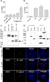
Figure 2. C. jejuni activates the inflammasome
(A–B) IL-1β secretion by primed J774.A1 macrophages infected (12 h) with different numbers of (A) C. jejuni, C. jejuni lysate (~m.o.i. 20) or (B) with different C. jejunistrains (m.o.i. 200). Infection with an m.o.i. higher than 20 (p < 0.05) or with different strains (p < 0.01) increased IL-1β secretion. Values are presented as the mean ± SEM of 3 independent experiments performed in duplicate. (C) Western blot probed for active IL-1β (17 kDa) (arrow) in the supernatant of primed J774.A1 macrophages after infection (12 h) with C. jejuni strain 108 (m.o.i. 20 or 200), 108cheY::cat (m.o.i. 20), E. coli (m.o.i 20), or without bacteria (non-stimulated). Lanes were loaded with 50 µg of protein. (D) Western blot probed for the cleaved caspase-1 p20 fragment in the supernatant of non-primed or primed J774.A1 macrophages after infection (6 h) with C. jejuni strain 108 (m.o.i. 200), E. coli (m.o.i. 20) or without bacteria (non-stimulated). Lanes were loaded with 10 µg of protein. (E) Confocal microscopy showing ASC speck formation in primed J774.A1 macrophages after infection (2 h) with mCherry fluorescent C. jejuni strain 108 (m.o.i. 40) or E. coli (m.o.i. 20) (red). ASC foci were stained with anti-ASC antibody in combination with goat-anti-rabbit-Alexa fluor 488 (green). Cell surface was stained with WGA-Alexa fluor 633 (blue).
Figure 3
Figure 3. Cellular infection-induced activation of the inflammasome
(A–C) Induction of IL-1β secretion in primed J774.A1 macrophages (12 h) exposed to different C. jejuni mutants and their respective parent strains 108, 81–176, 81116 (m.o.i. 20). (D) Effect of C. jejuniinfection of primed J774.A1 macrophages (m.o.i. 20) on IL-1β secretion (open bars) (after 12 h) and bacterial viability (closed bars) (after 6 h) as measured via the luciferase reporter assay (RLU). The P values (A–D) for all mutant strains compared to the parent strain were not significant different except for the cheY::cat and cetA::cat (P<0.05). (E) Intracellular bacterial viability of several C. jejuni strains (m.o.i. 20) in primed J774.A1 macrophages (6 h) as measured via the luciferase reporter assay. Strain 81–176 was significantly (P<0.01) more present intracellular than strain 108. Values are the mean ± SEM of 3 independent experiments performed in duplicate.
Figure 4
Figure 4. Cell viability and intracellular survival after C. jejuni induced inflammasome activation
(A–C) Cytotoxicity and bacterial survival in infected primed J774.A1 macrophages. (A) LDH release from primed cells at 12 h of incubation with ATP (5 mM), Salmonella (m.o.i. 20), E. coli (m.o.i. 20), or several C. jejuni (m.o.i. 20) or mutants of strain 108. ATP (P<0.001), Salmonella(P<0.01), and E. coli (P<0.01) caused significant cytotoxicity. None of the C. jejuni strains induced cytotoxicity. (B) Propidium iodide (PI) uptake by primed macrophages incubated (12 h) with the indicated strains. Salmonella (P<0.001) caused a significant increase in fluorescence intensity (F.I.). The increase F.I. induced by C. jejuni cheY mutant was not statistically significant. (C) Intracellular survival of C. jejuni strain 108 and its cheY derivative (m.o.i. 20) in primed J774.A1 macrophages as measured via the luciferase reporter assay. Luciferase activity (RLU) after 6 h infection was set as 100% value. Values are the mean ± SEM of 3 independent experiments performed in triplicate.
Figure 5
Figure 5. C. jejuni activation of the NLRP3 inflammasome in primary mouse macrophages
(A) Secretion of IL-1β by non-primed (open bars) and primed (closed bars) BMMs incubated (12 h) with Salmonella(m.o.i. 2), C. jejuni (m.o.i. 20), or ATP (2.5 mM). ATP (P<0.05) and Salmonella (P<0.001) significantly increased IL-1β secretion; C. jejuni significantly increased IL-1β in non-primed cells only (P<0.001). (B) Similar assay but with primed BMMs isolated from the indicated knockout mice and incubated with Salmonella (open bars) (m.o.i. 2) or ATP (2.5 mM) (closed bars). IL-1β secretion was significantly lower in caspase-1−/−/11−/−, NLRC4−/− BMMs upon infection with Salmonella (P<0.05). Upon ATP stimulation, IL-1β secretion was significantly reduced in caspase-1−/−/11−/−, ASC−/−, or NLRP3−/− BMMs (P<0.05). (C) IL-1β secretion by non-primed BMMs from several knockout mice infected (12 h) with C. jejuni strain 108 (open bars) or 108cheY::cat (closed bars) (m.o.i. 20). Significant lower secretion was observed for caspase-1−/−/11−/− (P<0.001), ASC−/− (P<0.001), and NLRP3−/− (P<0.01) BMMs upon stimulation with both C. jejuni strains. Strain cheY::cat induced more IL-1β secretion (P<0.05) than the parent strain. (D) Secretion of IL-1β in non-primed BMMs infected (12 h) with C. jejuni strain 108, 108cheY::cat or 108cheY::cat+pcheY (m.o.i. 20) showing that complementation of the defective cheY restored the high IL-1β secretion induced by strain 108 (P<0.01) to parental levels (P>.0.05). (E) Confocal microscopy on BMMs (wt and NLRP3−/−) infected (1 h) with mCherry fluorescent (red) C. jejuni 108 and 108cheY::cat (m.o.i 200); Cells were counterstained with WGA-Alexa fluor 633 (green). (F) Quantification of the number of intracellular bacteria in the wild type (open bars) and NLRP3−/− (closed bars) BMMs showed no significant difference in the number of intracellular bacteria between the wt and NLRP3−/− BMMs. Strain 108cheY::cat was more invasive (P<0.001) than the parent strain in the BMMs. Values are the mean ± SEM of 3 independent experiments performed in duplicate.
Figure 6
Figure 6. C. jejuni activation of the NLRP3 inflammasome in human macrophages
(A–B) Secretion of IL1-β by PMA differentiated THP-1 (open bars) and THP-1 defNLRP3 (closed bars) cells infected with Salmonella (m.o.i. 2), E. coli (m.o.i. 20), or different C. jejuni strains (m.o.i. 20) (12 h). Salmonella (P<0.01), E. coli (P<0.01) and all C. jejuni strains (P<0.05) induced IL-1β secretion upon infection. Strain cheY::cat induced more IL-1β secretion (P<0.01) than the parent strain. (C–D) Confocal microscopy on PMA differentiated THP-1 cells infected (2 h) with mCherry positive C. jejuni strain 108 (m.o.i. 40), E. coli (m.o.i. 20) (red). Cell surface was stained with WGA-Alexa fluor 633 (blue). ASC foci (C) were stained with an anti-ASC antibody in combination with goat-anti-rabbit-Alexa fluor 488 (green). Active caspase-1 (D) was detected with FLICA (green) at 1 h of infection. (E) Western blot showing the presence of active (cleaved) IL-1β (17 kDa) in the supernatant of non-infected and C. jejuni strain 108 (m.o.i. 20) infected (12 h) PMA differentiated THP-1 cells. Lanes were loaded with 50 µg of protein. (F) Intracellular viability of C. jejuni mutants and parent strain in 6 h infected PMA differentiated THP-1 (open bars) and THP-1 defNLRP3 (closed bars) as measured with the bacterial luciferase reporter assay. No significant differences in RLU were measured between the THP-1 and THP-1 defNLRP3. Strain 108cheY::cat was more invasive (P<0.001) than the parent strain. (G–H) Intracellular survival (24 h) of C. jejuni strains 108 (G) or 108cheY::cat (H) in THP-1 (open bars) or THP-1 defNLRP3 (closed bars) cells (24 h) at different duration of infection. Luciferase activity at 6 h of infection was set as 100%. The decrease in intracellular C. jejuni (108 and 108cheY::cat) at 12 h of infection was statistically significant (P<0.05). Values are the mean ± SEM of 3 independent experiments performed in duplicate.

References

    1. Thornberry NA, Bull HG, Calaycay JR, Chapman KT, Howard AD, Kostura MJ, Miller DK, Molineaux SM, Weidner JR, Aunins J. A novel heterodimeric cysteine protease is required for interleukin-1 beta processing in monocytes. Nature. 1992;356:768–774. - PubMed
    1. Fernandes-Alnemri T, Wu J, Yu JW, Datta P, Miller B, Jankowski W, Rosenberg S, Zhang J, Alnemri ES. The pyroptosome: a supramolecular assembly of ASC dimers mediating inflammatory cell death via caspase-1 activation. Cell Death Differ. 2007;14:1590–1604. - PMC - PubMed
    1. Martinon F, Burns K, Tschopp J. The inflammasome: a molecular platform triggering activation of inflammatory caspases and processing of proIL-1beta. Mol. Cell. 2002;10:417–426. - PubMed
    1. Latz E, Xiao TS, Stutz A. Activation and regulation of the inflammasomes. Nat. Rev. Immunol. 2013;13:397–411. - PMC - PubMed
    1. Strowig T, Henao-Mejia J, Elinav E, Flavell R. Inflammasomes in health and disease. Nature. 2012;481:278–286. - PubMed

Publication types

MeSH terms