. 2014 Nov 1;193(9):4643-53.
doi: 10.4049/jimmunol.1401625. Epub 2014 Sep 29.

Innate inflammation induced by the 8-oxoguanine DNA glycosylase-1-KRAS-NF-κB pathway

Affiliations

Innate inflammation induced by the 8-oxoguanine DNA glycosylase-1-KRAS-NF-κB pathway

Leopoldo Aguilera-Aguirre et al. J Immunol. .

Abstract

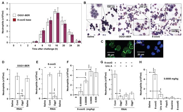
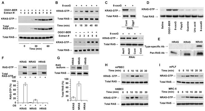
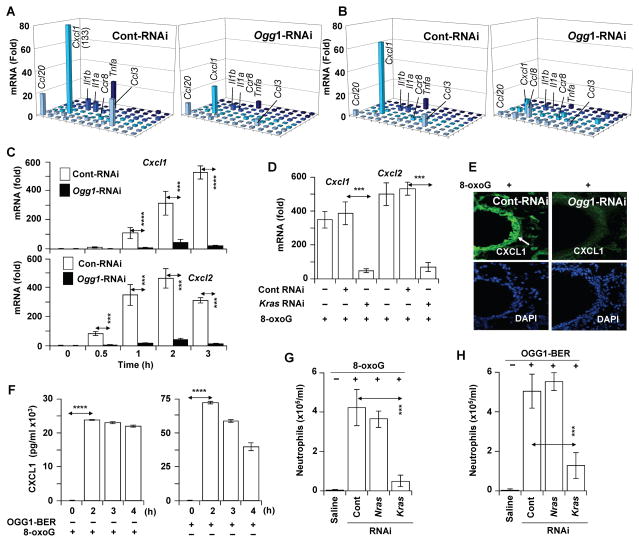
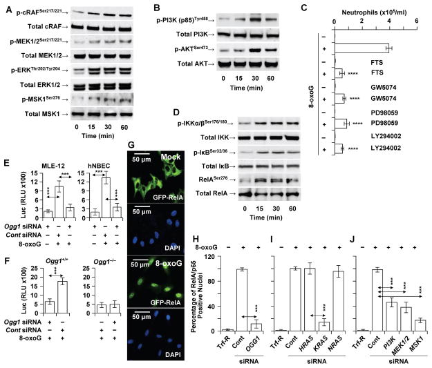
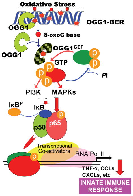
8-Oxoguanine-DNA glycosylase-1 (OGG1) is the primary enzyme for repairing 7,8-dihydro-8-oxoguanine (8-oxoG) via the DNA base excision repair pathway (OGG1-BER). Accumulation of 8-oxoG in the genomic DNA leads to genetic instability and carcinogenesis and is thought to contribute to the worsening of various inflammatory and disease processes. However, the disease mechanism is unknown. In this study, we proposed that the mechanistic link between OGG1-BER and proinflammatory gene expression is OGG1's guanine nucleotide exchange factor activity, acquired after interaction with the 8-oxoG base and consequent activation of the small GTPase RAS. To test this hypothesis, we used BALB/c mice expressing or deficient in OGG1 in their airway epithelium and various molecular biological approaches, including active RAS pulldown, reporter and Comet assays, small interfering RNA-mediated depletion of gene expression, quantitative RT-PCR, and immunoblotting. We report that the OGG1-initiated repair of oxidatively damaged DNA is a prerequisite for GDP → GTP exchange, KRAS-GTP-driven signaling via MAP kinases and PI3 kinases and mitogen-stress-related kinase-1 for NF-κB activation, proinflammatory chemokine/cytokine expression, and inflammatory cell recruitment to the airways. Mice deficient in OGG1-BER showed significantly decreased immune responses, whereas a lack of other Nei-like DNA glycosylases (i.e., NEIL1 and NEIL2) had no significant effect. These data unveil a previously unidentified role of OGG1-driven DNA BER in the generation of endogenous signals for inflammation in the innate signaling pathway.


Conflict of interest statement

Conflict of interest: The authors declare that no conflict of interest exists.

Figures

FIGURE 1
FIGURE 1
Experimental strategy and characterization of site-specific OGG1 depletion. (A) Experimental design. (B) Stealth RNAi decreased both Ogg1 mRNA and (C) protein levels in airway epithelial cells, as shown by qRT-PCR and immunohistochemistry, respectively. Green fluorescence mediated by Alexa-488 shows subcellular localization of OGG1 (left panels). Nuclei of cells were stained with DAPI (blue, right panels). (D) Changes in genomic 8-oxoG levels in OGG1-expressing (control RNAi; blue columns) and OGG1-depleted (Stealth RNAi) airway epithelial cells (red columns) as shown by FLARE Comet assays (Material and Methods). (E) Genomic 8-oxoG levels in the airway epithelium of OGG1-expressing mice at time 0 (upper panel) and 1h after oxidative challenge (lower panel) as shown by IHC (magnification x216). DAPI, 4′6-diamidino-2-phenylindole dihydrochloride; AE RNA, RNA isolated from exfoliated airway epithelial cells; RAS, mammalian homolog of viral H-, K-, N-RAS protein; MAPK(s), mitogen activated protein kinases; MSK1, mitogen- and stress-activated protein kinase1; PI3K, phosphoinositide 3-kinase; OGG1, 8-oxoguanine DNA glycosylase-1; 8-oxoG, 7,8-dihydro-8-oxoguanine; Trf-R, transfection reagent for in vivo use (Materials and Methods). *** = P<0.001
FIGURE 2
FIGURE 2
OGG1-initiated DNA base excision repair results in the recruitment of neutrophils. (A) Kinetics of neutrophil recruitment to the airways after OGG1-BER (empty columns) or challenge with 8-oxoG base (filled columns, 0.0005 mg/kg). Each time point represents the average number of neutrophils from 5 to 6 mice. (B) Representative images of differentially stained cells derived from the BALF of saline- (left panel), OGG1-BER (middle panel) and 8-oxoG (right panel)-challenged airways. (C) Immunohistochemical identification of neutrophils based on neutrophil elastase localized to cell membrane (Materials and Methods). Magnification x144. (D) OGG1 deficiency in airway epithelia decreases the accumulation of neutrophils. (E) The 8-oxoG base challenge-induced recruitment of neutrophils is OGG1-dependent. (F) 8-oxoG base induces dose-dependent increases in the recruitment of neutrophils to airways. (G) OGG1-independent changes in neutrophil numbers upon uric acid (0.0005 mg/kg) challenge. (H) 8-oxoG, but not 2,6-diamino-4-hydroxy-5-formamidopyrimidine (FapyG), 8-oxoguanine deoxynucleoside (8-oxodG) or guanine induces neutrophil recruitment. In A,D,F,G and H, BALF was obtained at 16 h post-challenge, total cell numbers were determined, and cyto-spin preparations made for differential staining with Wright-Giemsa to determine neutrophil counts. 8-oxoG, FapyG, 8-oxodG, guanine base or uric acid was introduced via the intranasal route in 60 μL saline. 8-oxoG, 7,8-dihydro-8-oxoguanine; OGG1-BER, OGG1-initiated DNA base excision repair; Uric A, uric acid; ** = P<0.01; *** = P<0.001; **** = P<0.0001.
FIGURE 3
FIGURE 3
OGG1-BER and its product 8-oxoG induce activation of KRAS. (A) OGG1-BER-induced activation of KRAS in the lungs as a function of time. At indicated times, lung extracts were prepared and changes in GTP-bound RAS were determined by active RAS pull-down assays. Subtype specific Ab to K-RAS and pan-RAS Ab was used to determine KRAS-GTP and total RAS levels, respectively (Materials and Methods). (B) KRAS activation after 8-oxoG challenge (time course, upper panels) and upon initiation of OGG1-BER (lower panels, 30 min time point is shown) in individual lungs. GTP-bound and total RAS levels were determined as described in legend to 3A. (C) Activation of KRAS is OGG1- (upper panel), but not NEIL1- or NEIL2-expression dependent (lower panel) in 8-oxoG-challenged airways. GTP-bound RAS levels were determined at 30 min post-challenge. (D), OGG1-BER and 8-oxoG challenge, but not FapyG, guanine, uric acid, or 8-oxodG activates RAS (KRAS) in the lungs. The 30 min time point is shown. GTP-bound RAS was determined as described in the legend to 3A. (E) Expression of NRAS, but not KRAS or HRAS in resident alveolar macrophages. Extracts of macrophages from unchallenged lungs (n = 6) were tested by WB using subtype-specific Abs. (F) GTP-bound RAS protein levels in pooled lung extracts (n = 5) after 8-oxoG challenge, and graphical depiction of the percentages of K-, N- and HRAS-GTP in total RAS-GTP. (G) Expression of RAS subtypes in lung. K-, N- and HRAS levels (upper panels) are shown by subtype-specific Abs. Lower panel shows graphical depiction of their percentages. Total RAS level is shown by pan-RAS Ab. In F and G, band intensities were quantified using Image J (ver. 1.44) software. Expression percentages were calculated using MS Excel. (H) KRAS activation in mouse (mPBEC) and human (hNBEC) bronchial airway epithelial cells upon 8-oxoG exposure. (I) Activation of N-RAS upon 8-oxoG exposure in mouse (mPLF) and human (MRC5) lung fibroblast cells. In A–I, 250 μg extract was utilized for assessing RAS-GTP levels; 20 μg for total RAS. In B (upper panel),C,D,F, and G, lungs were challenged with 60 μL saline containing 0.0005 mg per kg 8-oxoG. In H and I, cells were challenged with 10 μM of 8-oxoG solution in medium. In A and B (lower panels) oxidative DNA damage was generated by challenging the airways with glucose oxidase (1 mU in 60 μl saline). HRAS and KRAS, mammalian homolog of Harvey and Kirsten sarcoma virus oncogen; NRAS neuroblastoma RAS viral oncogene homolog; NEIL1 and 2, Nei (endonuclease VIII)-like DNA glycosylase-1 and -2; mPBEC, mouse primary bronchial epithelial cells; mPLF, mouse primary fibroblasts; hNBEC, human normal bronchial epithelial cells; MRC5, human diploid lung fibroblasts. FapyG, 2,6-diamino-4-hydroxy-5-formamidopyrimidine; 8-oxodG, 8-oxoguanine deoxynucleoside; 8-oxoG, 7,8-dihydro-8-oxoguanine.
FIGURE 4
FIGURE 4
OGG1-BER-dependent expression of proinflammatory chemokines and cytokines. (A,B) Expression of proinflammatory mediators upon OGG1-initiated DNA base excision repair (A) and after 8-oxoG (B) challenge of OGG1-expressing (Cont-RNAi, left panel) or OGG1-deficient (Ogg1-RNAi, right panel) lungs. RNAs were isolated at from five lungs and pooled, and the synthesized cDNAs were analyzed by Mouse Inflammatory Cytokines & Receptors PCR Array (2h time point is shown). (C) Time course of OGG1-dependent expression of Cxcl1 (upper panel) and Cxcl2 (lower panel) mRNAs upon 8-oxoG challenge (n=5–6; □, Cont-RNAi; ■, Ogg1-RNAi). (D) KRAS-dependent expression of Cxcl1 and Cxcl2 after 8-oxoG challenge to the airways. KRas was depleted from the airways by RNAi. RNAs isolated after 8-oxoG challenge were analyzed by qRT-PCR (n=5–6, 2 h time point is shown). (E) Expression of CXCL1 in the airway epithelia (white arrow) of OGG1-proficient (Cont-RNAi) and OGG1-deficient airways (Ogg1-RNAi), as shown by IHC. Representative lung sections (upper panels) are shown after a 2h 8-oxoG challenge (DAPI-staining of sections is shown on lower panel). (F) Levels of CXCL1 in the BALF of mice (n=5) challenged with 8-oxoG (left panel) or in response to OGG1-BER (n=6) as determined by BioPlex assays. (G) KRAS-, but not NRAS-expression-dependent recruitment of neutrophils to airways. RAS was depleted by RNAi to Kras or Nras, and airways (n=5–6) were challenged with 8-oxoG. Neutrophil numbers were determined 16 h post-challenge. (H) OGG1-BER-induced recruitment of neutrophils to airways is KRAS-, but not NRAS-expression dependent. RAS were depleted in airways (n=5–6) as in the legend to G, and neutrophil numbers were determined 16 h post-challenge. (B,C,D,F,G) Lungs were challenged with 60 μL saline containing 0.0005 mg per kg 8-oxoG. (A,F,H) Oxidative DNA damage was generated by challenging the airways with glucose oxidase (Materials and Methods). *** = P<0.001). Cxcl1, chemokine (C-X-C motif) ligand 1 transcript; Cxcl2, chemokine (C-X-C motif) ligand 2 transcript; Ccl20, C-C motif chemokine 20; Ccl3, chemokine (C-C motif) ligand 3; Il1a, Interleukin-1 alpha; Il1b, Interleukin-1 beta; Tnfa, tumor necrosis factor; CXCL1, chemokine (C-X-C motif) ligand 1. *** = P<0.001; **** = P<0.0001.
FIGURE 5
FIGURE 5
OGG1-BER-dependent activation of MAP, PI3 and MS kinases and NF-κB. (A,B) 8-oxoG challenge to the airways increased the phosphorylated levels of RAS targets, (A) MAP kinases (RAF1, MEK1/2, ERK1/2, and MSK1) and (B) PI3K, and AKT. In A,B, mice were challenged via the nasal route with 8-oxoG (0,0005 mg per kg), and the lungs were harvested after 0, 15, 30 and 60 min. Lung extracts were prepared from 5 animals per time points. Then 40 μg protein extract was fractionated by 5 to 20% SDS PAGE, and proteins transferred to membranes were identified by phopspho-specific and pan Abs (Materials and Methods). (C) Pharmacological inhibitors of RAS, RAF-1, MEK1/2 and PI3K decreased the recruitment of neutrophils to the airways. BALF was derived at 16 h post-challenge (n=6) and cell types determined by differential staining of cells. (D) Phosphorylation of IKK, I-κB and Rel/p65 in 8-oxoG-challenged airways. Mice were challenged, lung extracts were made and changes in phosphorylated and total IKK, I-κB and Rel/p65 was detected by immune-blotting using phopspho-specific and pan Abs as in legend to 5A,B. (E) OGG1-dependent activation from an NF-κB-driven promoter. MLE-12 and hNBEC were transfected with control or siRNA to OGG1, and 36 h later a luc2P reporter driven by NF-κB response elements was introduced and the cells exposed to 8-oxoG (10 μM). Luc activities were assessed 6 h later. (F) Ogg1+/+ and Ogg1−/− murine fibroblasts were transfected with control or siRNA to OGG1, and 36 h later, a luc2P reporter driven by NF-κB response elements was introduced and the cells exposed to 8-oxoG (10 μM). Luc activities were assessed 6 h later. (G) Nuclear translocation of GFP-RelA in A549 cells after exposure to 8-oxoG (10 μM). (H) Ablation of OGG1 or (I) KRAS by siRNA from GFP-RelA-expressing A549 cells decreased the 8-oxoG-induced nuclear accumulation of GFP-RelA. Ablation of H- and NRAS, had no effect. (J) siRNA depletion of PI3 or MAP kinases from GFP-RelA-expressing cells decreased the nuclear accumulation of GFP-RelA. RAF1, v-raf1 murine leukemia viral oncogene homolog 1; ERK1/2, extracellular signal-regulated kinase ½; MSK1, mitogen-stress related kinase-1; PI3K, phosphatidylinositol-4,5-bisphosphate 3-kinase; AKT, serine/threonine kinase (protein kinase B); KRAS, Kirsten rat sarcoma viral oncogene homolog; HRAS, Harvey rat sarcoma viral oncogene homolog; NRAS, neuroblastoma RAS viral oncogene homolog; GW5074, RAF-1 kinase inhibitor; PD98059, MAP kinase kinase (MEK1,2); LY294002, phosphatidylinositol 3-kinase inhibitor; FTS, trans-farnesyl-thiosalicylic acid (RAS membrane anchorage inhibitor). *** = P<0.01; **** = P< 0.001.
Figure 6
Figure 6
Schematic depiction of the role of OGG1-initiated DNA base excision repair in an innate immune response.

References

    1. D’Autreaux B, Toledano MB. ROS as signalling molecules: mechanisms that generate specificity in ROS homeostasis. Nat Rev Mol Cell Biol. 2007;8:813–824. - PubMed
    1. Dizdaroglu M. Oxidative damage to DNA in mammalian chromatin. Mutat Res. 1992;275:331–342. - PubMed
    1. Steenken S, Jovanovic SV. How easily oxidizable is DNA? One-electron reduction potentials of adenosine and guanosine radicals in aqueous solution. J Am Chem Soc. 1997;119:617–618.
    1. Chan SW, Dedon PC. The biological and metabolic fates of endogenous DNA damage products. J Nucleic Acids. 2010;2010:929047. - PMC - PubMed
    1. Shigenaga MK, Hagen TM, Ames BN. Oxidative damage and mitochondrial decay in aging. Proc Natl Acad Sci U S A. 1994;91:10771–10778. - PMC - PubMed

Publication types

MeSH terms

Associated data