. 2014 Sep 29;30(6):717-30.
doi: 10.1016/j.devcel.2014.08.003.

The architecture of CCAN proteins creates a structural integrity to resist spindle forces and achieve proper Intrakinetochore stretch

Affiliations

The architecture of CCAN proteins creates a structural integrity to resist spindle forces and achieve proper Intrakinetochore stretch

Aussie Suzuki et al. Dev Cell. .

Abstract

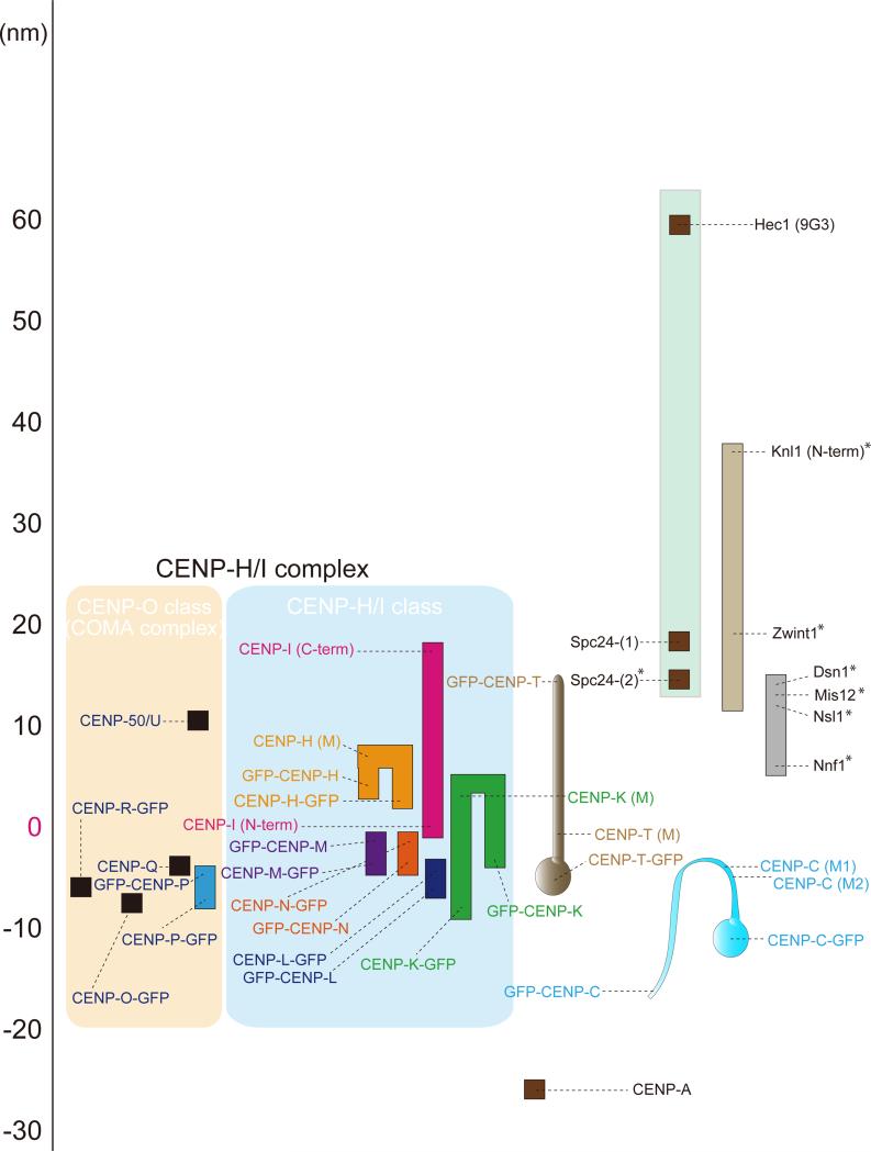
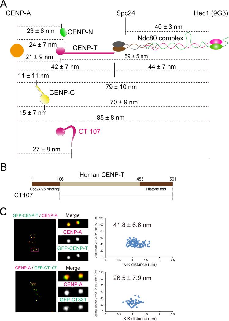
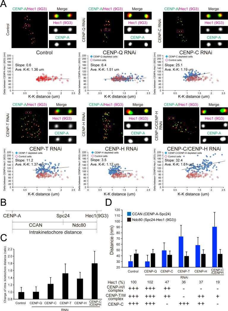
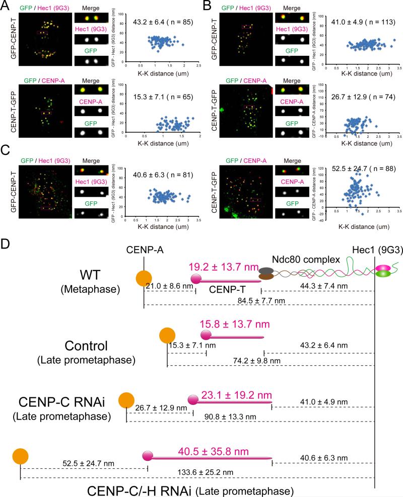
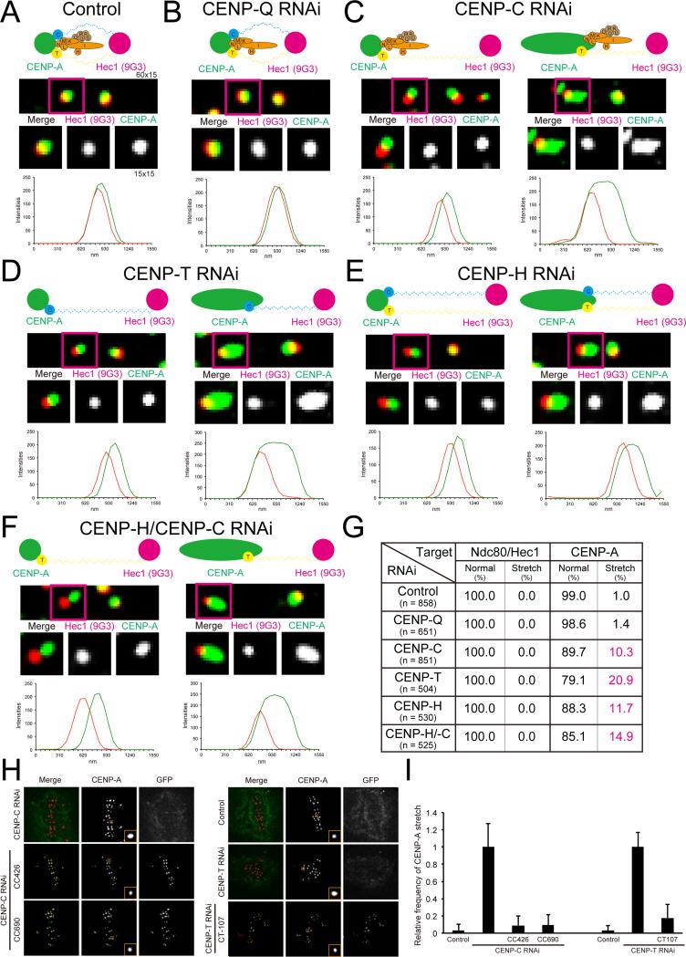
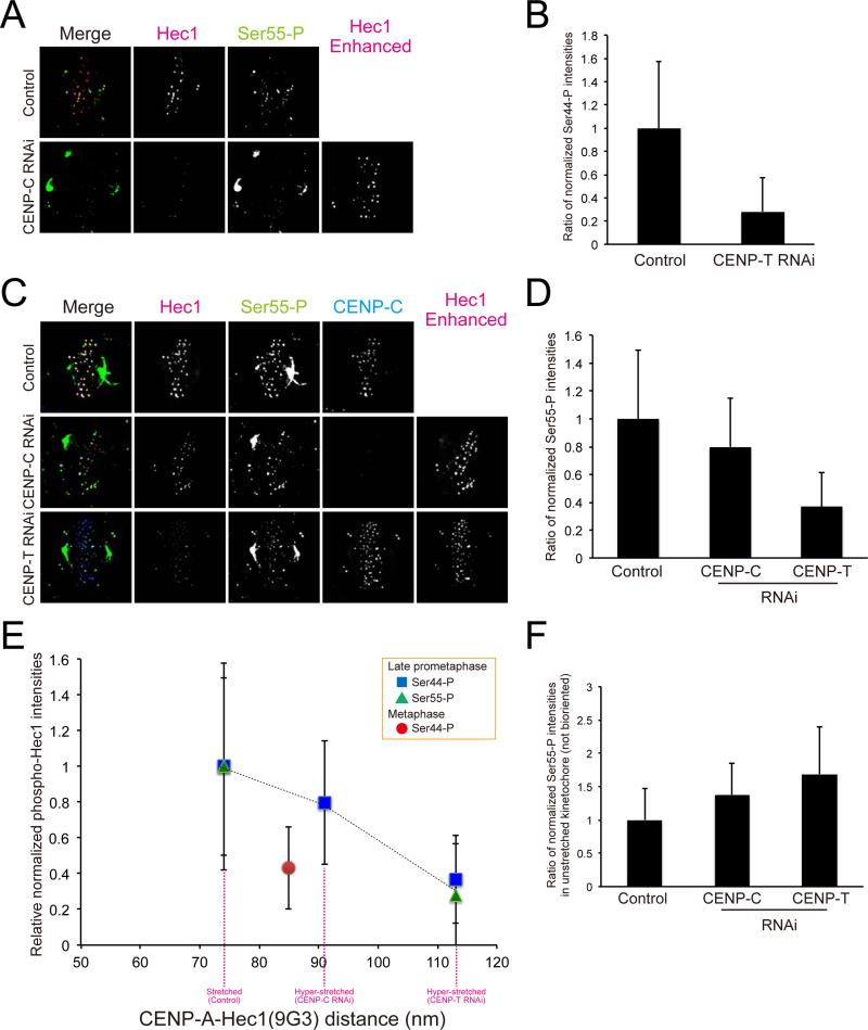
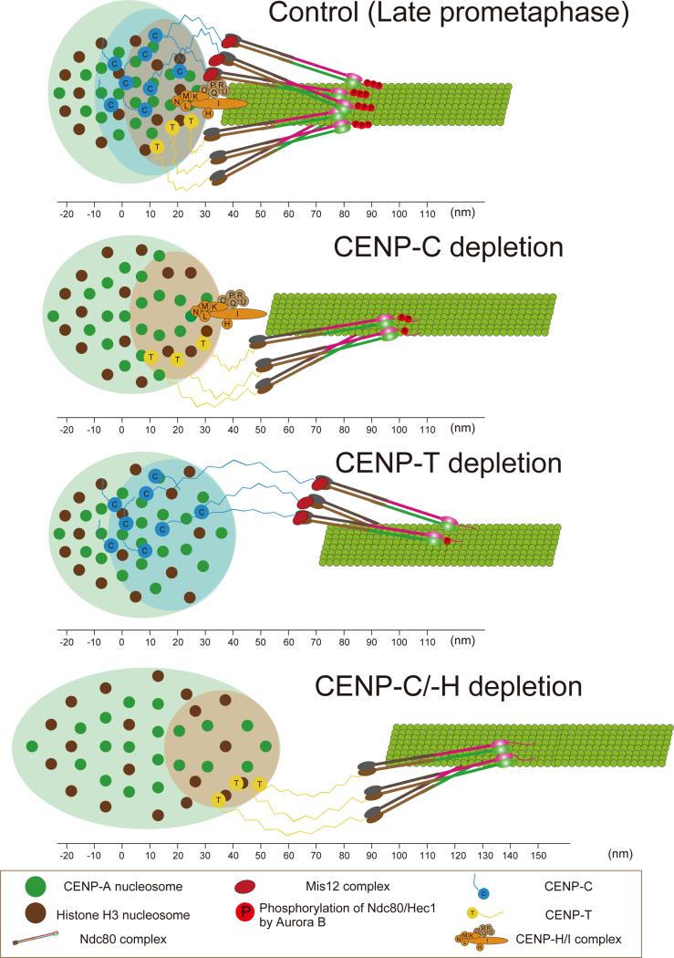
Constitutive centromere-associated network (CCAN) proteins, particularly CENP-C, CENP-T, and the CENP-H/-I complex, mechanically link CENP-A-containing centromeric chromatin within the inner kinetochore to outer kinetochore proteins, such as the Ndc80 complex, that bind kinetochore microtubules. Accuracy of chromosome segregation depends critically upon Aurora B phosphorylation of Ndc80/Hec1. To determine how CCAN protein architecture mechanically constrains intrakinetochore stretch between CENP-A and Ndc80/Hec1 for proper Ndc80/Hec1 phosphorylation, we used super-resolution fluorescence microscopy and selective protein depletion. We found that at bi-oriented chromosomes in late prometaphase cells, CENP-T is stretched ∼16 nm to the inner end of Ndc80/Hec1, much less than expected for full-length CENP-T. Depletion of various CCAN linker proteins induced hyper-intrakinetochore stretch (an additional 20-60 nm) with corresponding significant decreases in Aurora B phosphorylation of Ndc80/Hec1. Thus, proper intrakinetochore stretch is required for normal kinetochore function and depends critically on all the CCAN mechanical linkers to the Ndc80 complex.


Figures

Figure 1
Figure 1
Nanometer (nm) scale map of CCAN protein positions along the sister kinetochore axis relative to the KMN network proteins in human kinetochores at metaphase. The map was constructed from Delta measurements relative to Ndc80/Hec1(9G3) (Table S1 and Figures S1-S2). Scale on the far-left is set equal to zero at the position of the N-terminus of CENP-I centroid. Positive values are outward (towards the spindle microtubules), while negative values are inward (towards the centromere interior). Colored boxes indicate kinetochore proteins. Black dots indicate the mean Delta values that have been corrected for kinetochore tilt (Wan et al., 2009). The S.D. was typically 3-7 nm and, n > = 100 kinetochores averaged (Table S1). *Mean values obtained from Wan et al., 2009 and Varma et al., 2013.
Figure 2
Figure 2
Nanometer (nm) separations measured between mean centroid positions of key CCAN proteins in the linkage between CENP-A chromatin and the Ndc80 complex at metaphase. (A) Summary of separation measurements between the DNA binding domains of CENP-T, CENP-C, and CENP-N, within human kinetochores at metaphase relative to fluorescent labels on CENP-A and the Ndc80/Hec1 complex obtained from Delta analysis in Figure S2. Included are mean positions of the two ends of CT 107, which lacks the N-terminal of CENP-T that binds Spc24/25, as well as the positions of the two ends of normal CENP-T. Note that the mean position of the N-terminus of CT107 is close to the DNA binding C-terminal of CENP-T and not close to Spc24/Spc25. (B) Schematic representation of human CENP-T. The N-terminal region (1-106 for CENP-T) binds to Spc24/25 globular domain. The C-terminal region (455-561) is a histone fold domain for DNA binding. (C) Two-color immunofluorescence (left) and Delta analysis (right) in GFP-CENP-T (upper) and GFP-CT 107 (lower) expressed in CENP-T depleted cells.
Figure 3
Figure 3
CENP-T, CENP-C and CENP-H/I complex are needed to limit intrakinetochore stretch of bi-oriented chromosomes in late prometaphase. (A) Immunofluorescence of both CENP-A and Ndc80/Hec1 (9G3) at kinetochores in control cells or CENP-Q RNAi cells or CENP-C RNAi cells or CENP-T RNAi cells or CENP-H RNAi cells or CENP-C/CENP-H RNAi cells (upper). Distribution of Delta mean separation between CENP-A and Hec1 (9G3) in each cell (lower). (B) Diagram showing that total intrakinetochore distance between CENP-A and Ndc80/Hec1 (9G3) is the sum of the separation between CENP-A to the inner end of the Ndc80 complex (Spc24/Spc25) and length of the Ndc80 complex. (C) Intrakinetochore distance between CENP-A and Ndc80/Hec1(9G3) for control and experimental cells from mean Delta values in Figure 3A) normalized by the value for controls. (D) Intrakinetochore stretch of CCAN domain (CENP-A and Spc24) contributes to intrakinetochore stretch but length of the Ndc80 complex remains constant (separation between Spc24 to Ndc80/Hec1 (9G3)) in experimental cells compared to controls. All images in Figure 3 are corrected chromatic aberration.
Figure 4
Figure 4
Both CENP-A chromatin stretch and CENP-T stretch contribute to intrakinetochore stretch. Kinetochore immunofluorescence of both GFP and Ndc80/Hec1(9G3) or GFP and CENP-A in GFP-CENP-T or CENP-T-GFP stably expressed cell (A) treated with CENP-C RNAi (B) or CENP-C/-H RNAi (C). Corresponding Delta analysis (right). (D) Summary of mean separation measurements between CENP-A and C-terminus of CENP-T and the C-terminus of CENP-T to Spc24/Spc25 for changes in intrakinetochore stretch between normal late prometaphase and metaphase, and for changes in late prometaphase induced by depletion of CENP-C and CENP-C/-H.
Figure 5
Figure 5
CCAN regulates compaction of CENP-A containing chromatin during mitosis. (A) Two-color immunofluorescence of kinetochore CENP-A and Ndc80/Hec1(9G3) in control cells (top) with high magnification image of indicated region (middle), and line scan (bottom) for (A) Control; (B) CENP-Q depleted cells; (C) CENP-C depleted cells; (D) CENP-T depleted cells; (E) CENP-H depleted cells; and (F) CENP-C/CENP-H depleted cells. (G) The percentage of punctate spot or stretched spot of CENP-A and Ndc80/Hec1(9G3) immunofluorescence in control and experimental cells in (A) to (F). (H) Two-color immunofluorescence of CENP-A and GFP in control cells, CENP-C depleted cells, CENP-C depleted cells expressing GFP-CC-426 or GFP-CC-690, and CENP-T depleted cells expressing GFP-CC-426 or GFP-CT-107. (I) The relative frequency of CENP-A containing chromatin expansion for cells in (H).
Figure 6
Figure 6
Hyper-intrakinetochore stretch reduces the level of phosphorylation of the Ndc80/Hec1 N-terminus. (A) Two-color immunofluorescence for Ndc80/Hec1(9G3) and Ser44-P at kinetochores in control or CENP-T depleted cells. (B) Integrated fluorescence intensity of Ser44-P at kinetochores normalized by the value for Ndc80/Hec1(9G3) on bi-oriented kinetochores in control or CENP-T depleted cells. (C) Two-color immunofluorescence forNdc80/Hec1(9G3) and Ser55-P at kinetochores in CENP-C depleted or CENP-T depleted cells. (D) Integrated fluorescence intensity of Ser55-P at kinetochores normalized by the value for Ndc80/Hec1(9G3) on bi-oriented kinetochores in control, CENP-C depleted, and CENP-T depleted cells. (E) Data from (B) and (D) plotted as a function of the separation between CENP-A to Ndc80/Hec1. (F) Integrated fluorescence intensity of Ser55-P at kinetochores normalized by the value for Ndc80/Hec1(9G3) on unaligned kinetochores in control, CENP-C depleted, and CENP-T depleted cells. The enhanced Hec1(9G3) images are added to mark kinetochore positions in (A) and (B).
Figure 7
Figure 7
A model for inner kinetochore protein architecture and how phosphorylation and the N-terminal kMT binding domain of the Ndc80 complex depends on intrakinetochore stretch for bi-oriented centromeres late prometaphase and metaphase control cells (Top panel) and in late prometaphase for CENP-C depleted, CENP-T depleted, and CENP-C/-H depleted human cells (Bottom 3 panels) . From the periphery of the centromeric chromatin, the depth of CENP-A is greatest, with the DNA binding domains of CENP-C being slightly deeper than those of CENP-T and CENP-N. CENP-T/-W/-S/-X complex, CENP-C, and CENP-H/I complex all contribute to limiting the separation between the mean positions of CENP-A and the inner end of the Ndc80 complex to about 40 nm for bi-oriented centromeres in control cells. This separation is increased to, much less than is possible and contribute for kMT assembly regulated by Aurora B. The depletion of CENP-T or CENP-C induced hyper-intrakinetochore stretch and a reduction of phosphorylation of Hec1 by Aurora B.

References

    1. Amano M, Suzuki A, Hori T, Backer C, Okawa K, Cheeseman IM, Fukagawa T. The CENP-S complex is essential for the stable assembly of outer kinetochore structure. The Journal of cell biology. 2009;186:173–182. - PMC - PubMed
    1. Bakhoum SF, Thompson SL, Manning AL, Compton DA. Genome stability is ensured by temporal control of kinetochore-microtubule dynamics. Nature cell biology. 2009;11:27–35. - PMC - PubMed
    1. Biggins S. The composition, functions, and regulation of the budding yeast kinetochore. Genetics. 2013;194:817–846. - PMC - PubMed
    1. Campbell CS, Desai A. Tension sensing by Aurora B kinase is independent of survivin-based centromere localization. Nature. 2013;497:118–121. - PMC - PubMed
    1. Carroll CW, Milks KJ, Straight AF. Dual recognition of CENP-A nucleosomes is required for centromere assembly. The Journal of cell biology. 2010;189:1143–1155. - PMC - PubMed

Publication types

LinkOut - more resources