

Corneal blindness and xenotransplantation
- PMID: 25268248
- PMCID: PMC4181387
- DOI: 10.1111/xen.12082
Corneal blindness and xenotransplantation
Abstract
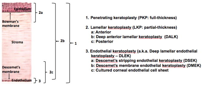
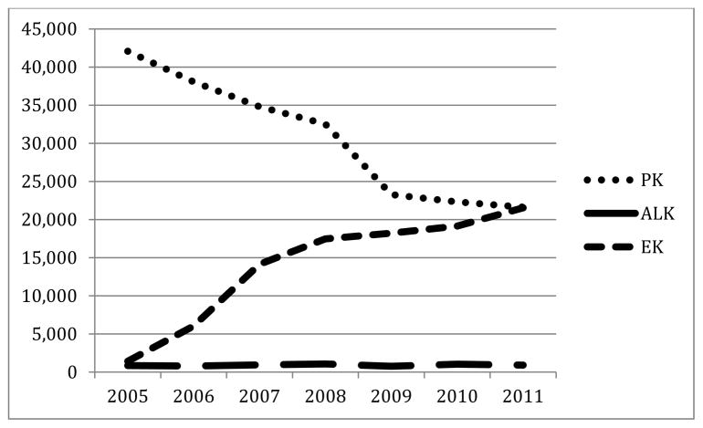
Approximately 39 million people are blind worldwide, with an estimated 285 million visually impaired. The developing world shoulders 90% of the world's blindness, with 80% of causative diseases being preventable or treatable. Blindness has a major detrimental impact on the patient, community, and healthcare spending. Corneal diseases are significant causes of blindness, affecting at least 4 million people worldwide. The prevalence of corneal disease varies between parts of the world. Trachoma, for instance, is the second leading cause of blindness in Africa, after cataracts, but is rarely found today in developed nations. When preventive strategies have failed, corneal transplantation is the most effective treatment for advanced corneal disease. The major surgical techniques for corneal transplantation include penetrating keratoplasty (PK), anterior lamellar keratoplasty, and endothelial keratoplasty (EK). Indications for corneal transplantation vary between countries, with Fuchs' dystrophy being the leading indication in the USA and keratoconus in Australia. With the exception of the USA, where EK will soon overtake PK as the most common surgical procedure, PK is the overwhelming procedure of choice. Success using corneal grafts in developing nations, such as Nepal, demonstrates the feasibility of corneal transplantation on a global scale. The number of suitable corneas from deceased human donors that becomes available will never be sufficient, and so research into various alternatives, for example stem cells, amniotic membrane transplantation, synthetic and biosynthetic corneas, and xenotransplantation, is progressing. While each of these has potential, we suggest that xenotransplantation holds the greatest potential for a corneal replacement. With the increasing availability of genetically engineered pigs, pig corneas may alleviate the global shortage of corneas in the near future.
Keywords: corneal transplantation; corneas; developing world; donor shortage; keratoplasty; pig; xenotransplantation.
© 2014 John Wiley & Sons A/S.
Conflict of interest statement
No author reports a conflict of interest.
Figures
References
-
- THURSTON M, THURSTON A, MCLEOD J. Socio-emotional effects of the transition from sight to blindness. Br J Vis Impair. 2010;28:90–112.
-
- TAN DT, DART JK, HOLLAND EJ, et al. Corneal transplantation. Lancet. 2012;379:1749–1761. - PubMed
-
- HARA H, COOPER DKC. The immunology of corneal xenotransplantation: a review of the literature. Xenotransplantation. 2010;17:338–349. - PubMed
-
- KIM MK, WE WR, PARK CG, et al. Xenocorneal transplantation. Curr Opin Organ Transplant. 2011;16:231–236. - PubMed
Publication types
MeSH terms
Grants and funding
LinkOut - more resources
Full Text Sources
Other Literature Sources
Medical
Research Materials
