. 2014 Oct 28;53(42):6628-40.
doi: 10.1021/bi5005552. Epub 2014 Oct 14.

Covalent small molecule inhibitors of Ca(2+)-bound S100B

Affiliations

Covalent small molecule inhibitors of Ca(2+)-bound S100B

Michael C Cavalier et al. Biochemistry. .

Abstract

Elevated levels of the tumor marker S100B are observed in malignant melanoma, and this EF-hand-containing protein was shown to directly bind wild-type (wt) p53 in a Ca(2+)-dependent manner, dissociate the p53 tetramer, and inhibit its tumor suppression functions. Likewise, inhibiting S100B with small interfering RNA (siRNA(S100B)) is sufficient to restore wild-type p53 levels and its downstream gene products and induce the arrest of cell growth and UV-dependent apoptosis in malignant melanoma. Therefore, it is a goal to develop S100B inhibitors (SBiXs) that inhibit the S100B-p53 complex and restore active p53 in this deadly cancer. Using a structure-activity relationship by nuclear magnetic resonance approach (SAR by NMR), three persistent binding pockets are found on S100B, termed sites 1-3. While inhibitors that simultaneously bind sites 2 and 3 are in place, no molecules that simultaneously bind all three persistent sites are available. For this purpose, Cys84 was used in this study as a potential means to bridge sites 1 and 2 because it is located in a small crevice between these two deeper pockets on the protein. Using a fluorescence polarization competition assay, several Cys84-modified S100B complexes were identified and examined further. For five such SBiX-S100B complexes, crystallographic structures confirmed their covalent binding to Cys84 near site 2 and thus present straightforward chemical biology strategies for bridging sites 1 and 3. Importantly, one such compound, SC1982, showed an S100B-dependent death response in assays with WM115 malignant melanoma cells, so it will be particularly useful for the design of SBiX molecules with improved affinity and specificity.


Figures

Figure 1
Figure 1
Binding sites 1–3. Shown is a ribbon diagram of the S100B dimer with the three persistent binding sites shaded. The sites were identified in CaS100B–target and CaS100B–SBiX complexes. Site 1 interactions were first highlighted via the structure of CaS100B bound to the C-terminal regulatory domain of p53, while sites 2 and 3 were elucidated in the detailed characterization of the CaS100B–SBi1 complex.
Figure 2
Figure 2
Assigned two-dimensional 1H–15N HSQC NMR spectra. The binding of five small molecules (SC124, SC1475, SC1982, SBi4172, and SBi4434) to CaS100B was assessed by monitoring perturbations of backbone 1H–15N HSQC NMR experiments. Chemical shift perturbations in NMR spectra for CaS100B in the presence of SBi4172 could not be measured as the compound caused CaS100B to from soluble aggregates. The solid horizontal line is plotted at the mean perturbation (in hertz) plus one standard deviation for each data set.
Figure 3
Figure 3
Chemical modification of Cys84 of CaS100B. Shown are schematics that provide an overview of the chemical modification of Cys84 by compounds (A) SC124, (B) SC1475, (C) SC1982, (D) SBi4172, and (E) SBi4434.
Figure 4
Figure 4
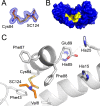
Crystallographic structure of the CaS100B–SC124 complex. (A) Shown is an |Fo| – |Fc| electron density omit map of SC124 covalently bound to Cys84 of CaS100B with the map contoured at the 2.5σ level. (B) Dimeric CaS100B is rendered in a surface diagram with residues within 4 Å of the SC124 colored yellow. SC124 is found within persistent binding site 2. (C) SC124 is situated within the hydrophobic pocket formed by Val8, Phe43, Phe87, and Phe88. SC124 makes no hydrogen bonds. The neighboring Zn2+ binding site composed of Glu89, His15, His25, and His85 is also rendered.
Figure 5
Figure 5
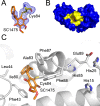
Crystallographic structure of the CaS100B–SC1475 complex. (A) Shown is an |Fo| – |Fc| electron density omit map of SC1475 covalently bound to Cys84 of CaS100B with the map contoured at the 2.5σ level. (B) Dimeric CaS100B is rendered in a surface diagram with residues within 4 Å of the SC1475 colored yellow. SC1475 is found within persistent binding site 2. (C) SC1475 is situated within the hydrophobic pocket formed by Phe43, Leu44, Ile80, Ala83, Phe87, and Phe88. SC1475 makes no hydrogen bonds. The neighboring Zn2+ binding site composed of Glu89, His15, His25, and His85 is also rendered.
Figure 6
Figure 6
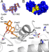
Crystallographic structure of the CaS100B–SC1982 complex. (A) Shown is an |Fo| – |Fc| electron density omit map of SC1982 covalently bound to Cys84 of CaS100B with the map contoured at the 2.5σ level. (B) Dimeric CaS100B is rendered in a surface diagram with residues within 4 Å of the SC1982 colored yellow. SC1982 is found within persistent binding site 2. (C) SC1982 is situated within the hydrophobic pocket formed by Phe43, Ala83, Phe87, and Phe88. SC1982 makes hydrogen bonds to the backbone carbonyls of His42 and Phe43. The neighboring Zn2+ binding site composed of Glu89, His15, His25, and His85 is also rendered.
Figure 7
Figure 7
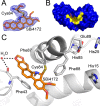
Crystallographic structure of the CaS100B–SBi4172 complex. (A) Shown is an |Fo| – |Fc| electron density omit map of SBi4172 covalently bound to Cys84 of CaS100B with the map contoured at the 2.5σ level. (B) Dimeric CaS100B is rendered in a surface diagram with residues within 4 Å of the SBi4172 colored yellow. SBi4172 is found within persistent binding site 2. (C) SBi4172 is situated within the hydrophobic pocket formed by Phe43, Phe87, and Phe88. SBi4172 makes a water-bridged interaction with the backbone carbonyl of Phe43. The neighboring Zn2+ binding site composed of Glu89, His15, His25, and His85 is also rendered.
Figure 8
Figure 8
Crystallographic structure of the CaS100B–SBi4434 complex. (A) Shown is an |Fo| – |Fc| electron density omit map of SBi4434 covalently bound to Cys84 of CaS100B with the map contoured at the 2.5σ level. (B) Dimeric CaS100B is rendered in a surface diagram with residues within 4 Å of the SBi4434 colored yellow. SBi4434 is found within persistent binding site 2. (C) SBi4434 is situated within the hydrophobic pocket formed by Phe43, Phe87, and Phe88. SBi4434 makes no hydrogen bonds. The neighboring Zn2+ binding site composed of Glu89, His15, His25, and His85 is also rendered.
Figure 9
Figure 9
Westen blot analysis. The control without drug was treated with the same DMSO concentration (0.01%). The addition of SC1982 did not affect the protein levels of S100B in the shRNAscrambled or shRNAS100B cell lines. However, both shRNAscrambled and shRNAS100B cell lines had a measurable gain in total p53 protein levels in the presence of SC1982. This result along with an observation of S100B expression-dependent growth inhibition suggests that SC1982 displays an on-target effect of S100B inhibition with subsequent p53 restoration.
Figure 10
Figure 10
Distortion of the zinc binding site is a hallmark of the S100B covalent inhibitor family. Shown are ribbon diagrams of CaS100B in the presence (yellow) and absence (gray) of a covalent modifier (SC1982) with residues and ligands of interest shown as sticks. The covalent modifier family of SBiXs is hallmarked by perturbation in the conformational state of the residues forming the Zn2+ binding site (His15, His25, His85, and Glu89). Commonly, Glu89 is seen intruding on the Zn2+ with a subsequent displacement of a histidine side chain.

References

    1. Kwong L. N.; Davies M. A. (2014) Targeted therapy for melanoma: Rational combinatorial approaches. Oncogene 33, 1–9. - PubMed
    1. Topalian S. L.; Hodi F. S.; Brahmer J. R.; Gettinger S. N.; Smith D. C.; McDermott D. F.; Powderly J. D.; Carvajal R. D.; Sosman J. A.; Atkins M. B.; Leming P. D.; Spigel D. R.; Antonia S. J.; Horn L.; Drake C. G.; Pardoll D. M.; Chen L.; Sharfman W. H.; Anders R. A.; Taube J. M.; McMiller T. L.; Xu H.; Korman A. J.; Jure-Kunkel M.; Agrawal S.; McDonald D.; Kollia G. D.; Gupta A.; Wigginton J. M.; Sznol M. (2012) Safety, activity, and immune correlates of anti-PD-1 antibody in cancer. N. Engl. J. Med. 366, 2443–2454. - PMC - PubMed
    1. Bollag G.; Hirth P.; Tsai J.; Zhang J.; Ibrahim P. N.; Cho H.; Spevak W.; Zhang C.; Zhang Y.; Habets G.; Burton E. A.; Wong B.; Tsang G.; West B. L.; Powell B.; Shellooe R.; Marimuthu A.; Nguyen H.; Zhang K. Y.; Artis D. R.; Schlessinger J.; Su F.; Higgins B.; Iyer R.; D’Andrea K.; Koehler A.; Stumm M.; Lin P. S.; Lee R. J.; Grippo J.; Puzanov I.; Kim K. B.; Ribas A.; McArthur G. A.; Sosman J. A.; Chapman P. B.; Flaherty K. T.; Xu X.; Nathanson K. L.; Nolop K. (2010) Clinical efficacy of a RAF inhibitor needs broad target blockade in BRAF-mutant melanoma. Nature 467, 596–599. - PMC - PubMed
    1. Flaherty K. T.; Puzanov I.; Kim K. B.; Ribas A.; McArthur G. A.; Sosman J. A.; O’Dwyer P. J.; Lee R. J.; Grippo J. F.; Nolop K.; Chapman P. B. (2010) Inhibition of mutated, activated BRAF in metastatic melanoma. N. Engl. J. Med. 363, 809–819. - PMC - PubMed
    1. Espinosa E.; Grob J. J.; Dummer R.; Rutkowski P.; Robert C.; Gogas H.; Kefford R.; Eggermont A. M.; Martin Algarra S.; Hauschild A.; Schadendorf D. (2014) Treatment Algorithms in Stage IV Melanoma. American Journal of Therapeutics DOI: 10.1097/MJT.0b013e31829e885c. - PubMed

Publication types

MeSH terms