. 2014 Sep 30;12(9):e1001957.
doi: 10.1371/journal.pbio.1001957. eCollection 2014 Sep.

New developmental evidence clarifies the evolution of wrist bones in the dinosaur-bird transition

Affiliations

New developmental evidence clarifies the evolution of wrist bones in the dinosaur-bird transition

João Francisco Botelho et al. PLoS Biol. .

Abstract

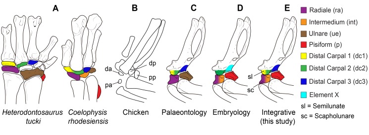
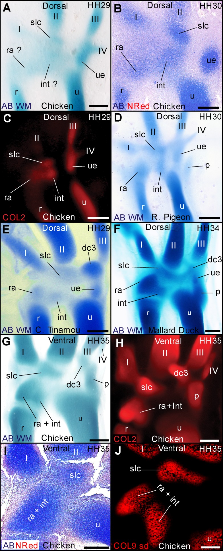
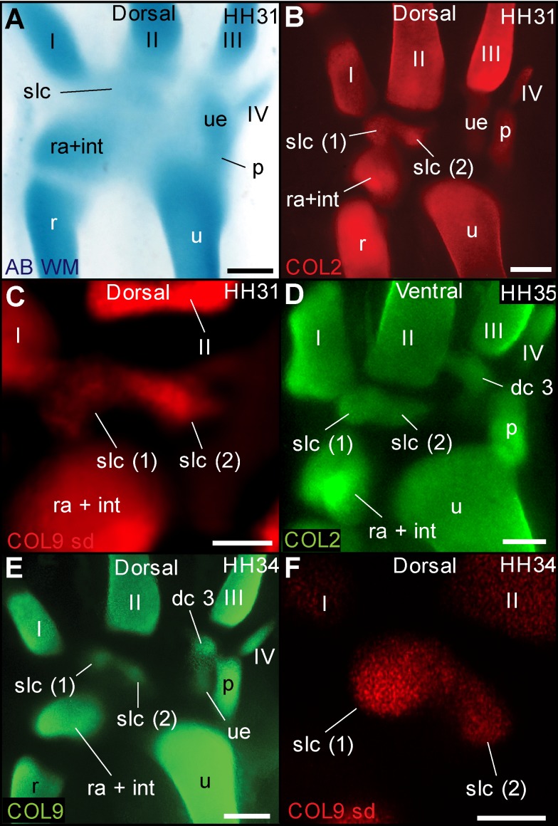
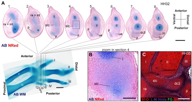
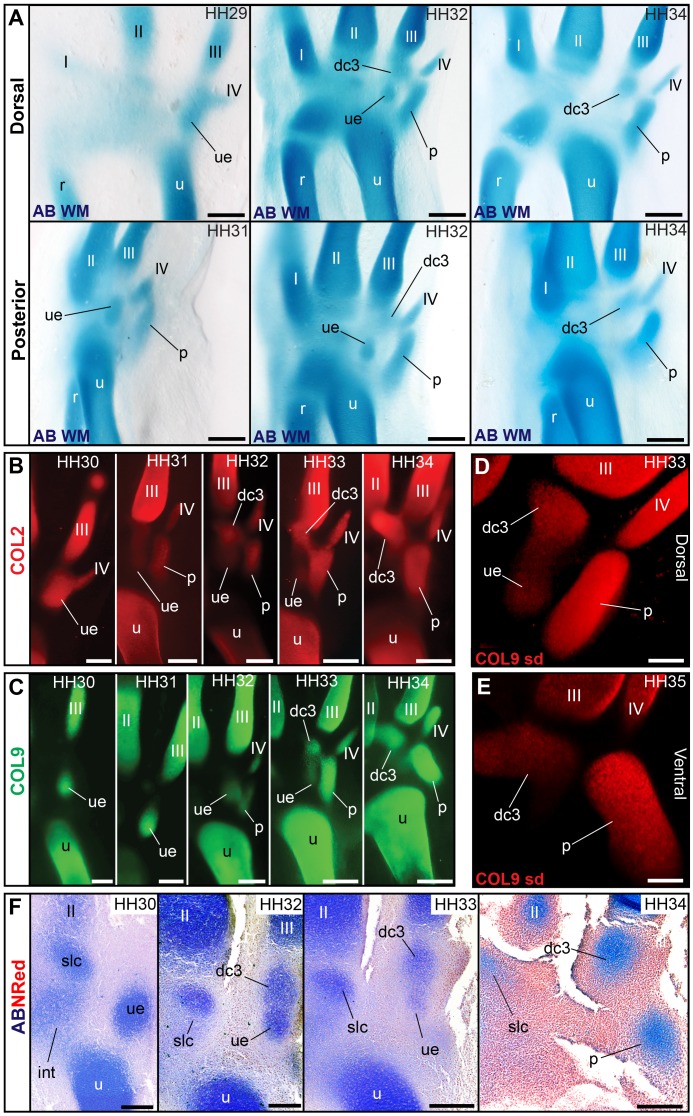
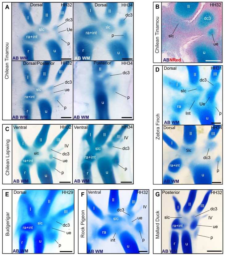
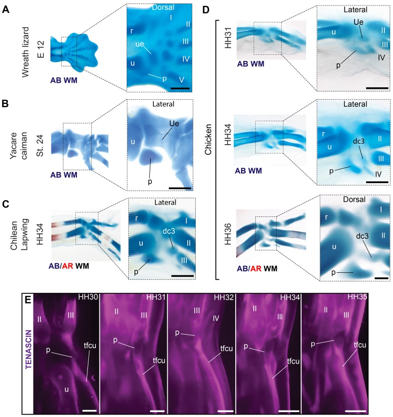
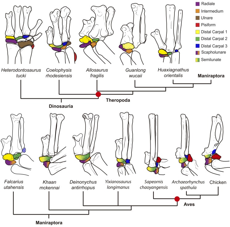
From early dinosaurs with as many as nine wrist bones, modern birds evolved to develop only four ossifications. Their identity is uncertain, with different labels used in palaeontology and developmental biology. We examined embryos of several species and studied chicken embryos in detail through a new technique allowing whole-mount immunofluorescence of the embryonic cartilaginous skeleton. Beyond previous controversy, we establish that the proximal-anterior ossification develops from a composite radiale+intermedium cartilage, consistent with fusion of radiale and intermedium observed in some theropod dinosaurs. Despite previous claims that the development of the distal-anterior ossification does not support the dinosaur-bird link, we found its embryonic precursor shows two distinct regions of both collagen type II and collagen type IX expression, resembling the composite semilunate bone of bird-like dinosaurs (distal carpal 1+distal carpal 2). The distal-posterior ossification develops from a cartilage referred to as "element x," but its position corresponds to distal carpal 3. The proximal-posterior ossification is perhaps most controversial: It is labelled as the ulnare in palaeontology, but we confirm the embryonic ulnare is lost during development. Re-examination of the fossil evidence reveals the ulnare was actually absent in bird-like dinosaurs. We confirm the proximal-posterior bone is a pisiform in terms of embryonic position and its development as a sesamoid associated to a tendon. However, the pisiform is absent in bird-like dinosaurs, which are known from several articulated specimens. The combined data provide compelling evidence of a remarkable evolutionary reversal: A large, ossified pisiform re-evolved in the lineage leading to birds, after a period in which it was either absent, nonossified, or very small, consistently escaping fossil preservation. The bird wrist provides a modern example of how developmental and paleontological data illuminate each other. Based on all available data, we introduce a new nomenclature for bird wrist ossifications.


Conflict of interest statement

The authors have declared that no competing interests exist.

Figures

Figure 1
Figure 1. Current hypotheses on the ossifications present in the wrist of birds.
(A) The carpal skeleton of early dinosaurs (Heterodontosaurus, Coelophysis). Colored elements represent bones that are potentially still present in the avian wrist. (B) The four carpal ossifications of birds as observed in the chicken at 21 d posthatching. The distal–anterior (da) and distal–posterior ossifications thereafter fuse to each other and to the metacarpi. The proximal–posterior (pp) and proximal–anterior (pa) remain unfused. (C) An identification of the four ossifications in the adult chicken wrist as often used in palaeontology. The proximal–posterior ossification is the ulnare (brown), the proximal–anterior ossification is the radiale (purple), the distal–anterior ossification is considered to be a composite of dcI+dcII (yellow+green), and the distal–posterior ossification is considered to be dcIII (dark blue). (D) An identification of the four ossifications in the avian wrist as often used in embryology. The proximo–posterior ossification is the pisiform (red), the proximo–anterior ossification is the radiale+intermedium (purple+orange), the distal–anterior ossification is DCII (green), and the distal–posterior ossification is a neomorphic “element x” (light blue). Despite these general trends, authors in either field may use a different combination of these nomenclatures. (E) Identification of the ossifications in the avian wrist according to the evidence discussed in the present work. We support the use of the term “scapholunare” for the bone that develops from the embryonic cartilage that is composite of radiale+intermedium, and “semilunate” for the ossification that develops from the embryonic cartilage that is a composite of Dc1+Dc2.
Figure 2
Figure 2. Phylogenetic relationships among the modern taxa used in this study.
Figure 3
Figure 3. Evidence for a composite radiale+intermedium cartilage in avian embryos.
(A) Alcian blue in the chicken shows diffuse staining along the anterior-mid region of the proximal carpus, providing no evidence for a separate radiale and intermedium. (B) Histological sections in the chicken, however, reveal two distinct cartilaginous foci. (C) Immunofluorescence for collagen type II also reveals two separate foci of early expression. (D) Alcian blue is sufficient to observe a separate radiale and intermedium in the development of the pigeon and (E) Chilean tinamou. At later stages, the bilobed shape of the proximal–anterior cartilage suggests it contains the radiale and intermedium in the duck (F) and in (G) the chicken. (I) Collagen type II immunoflourescence also reveals a bi-lobed shape. (H) A histological section of a late stage in the chicken reveals a single perichondrium, with no internal division or septum. (J) Despite this, two separate domains of collagen type IX expression are very distinct, as observed using spinning disc microscopy. These results confirm the composite nature (radiale+intermedium) of the cartilage that gives rise to the proximal–anterior ossification. Scale bars, (A, B, and F) 300 µm, (C) 400 µm, (D–I) 500 µm, and (J) 200 µm.
Figure 4
Figure 4. Two regions of collagen expression support the composite nature of the cartilage that becomes the distal–anterior ossification.
(A) Whole-mount alcian blue staining in the chicken and all species observed provides no evidence for separate cartilages in the diffusely stained region where the distal–anterior ossification will form (labelled slc; see also Figure 4A–B). However, (B) collagen type II and (C) collagen type IX in this region show two distinct regions of early expression. (D) Later, collagen type II expression becomes more continuous (see also Figure 4C), but collagen type IX expression (E) reveals two nearly separate regions, shown in detail in (F) using spin disc microscopy (see Video S1). Scale bars, (A) 300 µm, (B and D) 400 µm, (C) 200 µm, (E) 500 µm, and (F) 100 µm.
Figure 5
Figure 5. Traditional techniques for cartilage visualization in the region giving rise to the distal–anterior ossification.
(A) Stacks of anterior–posterior histological sections, with zoom-in to one section (B) revealing asymmetric tissue organization, with a concentric focus of cells and stronger alcian staining towards posterior. (C) A section in a dorso-ventral stack of a later stage reveals a well-defined cartilage (stained with safranin red) with a single perichondrium and no internal septum or separation. Scale bars, (A and B) 500 µm and (C) 1 mm.
Figure 6
Figure 6. Loss of the ulnare and late formation of distal carpal 3 (“element x”) in the chicken.
(A) Whole-mount alcian blue staining confirms the ulnare is the first carpal formed in avian embryos, distal to the ulna. Thereafter, a distal carpal 3 (referred to as “element x” in previous embryological descriptions) is formed distal to the ulnare, coexisting with it. Finally, the ulnare disappears, whereas dc3 persists. (B) Collagen type II and (C) collagen type IX whole-mount immunostaining documents the formation of dc3 distal to the ulnare and the reduction and disappearance of the ulnare. (D) Detail of dc3 and receding ulnare, coexisting in the chicken embryo, as observed by spin-disc microscopy. See Video S2. (E) Detail of dc3 after disappearance of the ulnare. The dc3 cartilage thereafter acquires a bent, “v”-like shape in galloanserae (chicken and duck), but not other bird species observed (Video S3). (F) Histological sections showing the late formation of dc3, its co-existence with the receding ulnare, and the disappearance of the ulnare in the chicken embryo. Scale bars, (A–C and F) 300 µm and (D and E) 150 µm.
Figure 7
Figure 7. Coexistence of dc3 and the ulnare in a diverse sample of avian taxa.
(A) Whole-mount alcian blue staining in the Chilean tinamou showing co-existence and subsequent disappearance of the ulnare. (B) Histological section in a dorso-ventral stack of the Chilean tinamou showing coexistence of the ulnare and dc3. (C) Whole-mount alcian blue staining showing coexistence of the ulnare and dc3 in the Chilean lapwing. (D) Coexistence of ulnare and dc3 and disappearance of the ulnare in zebra finch. (E) Coexistence of ulnare and dc3 in (E) budgerigar, (F) pigeon, and (G) duck. Scale bars, (A and C) 400 µm, (B) 200 µm, (C, G, and F) 500 µm, and (E–D) 300 µm.
Figure 8
Figure 8. The posterior–proximal ossification of the wing develops from the embryonic cartilage that corresponds to the pisiform of reptiles.
(A) The pisiform in embryos of the Wreath lizard and (B) a caiman demonstrates its typical position for amniotes, postero-ventral to the connection of the ulna and ulnare. The cartilage that gives rise to the proximal-posterior bone is found in a comparable position in birds, as shown for (C) Chilean lapwing and (D) a developmental series of chicken. (E) Immunofluorescence for tenascin shows the development of this cartilage is always associated to the tendon of the flexor carpi ulnaris muscle (tfcu), at the turn of the wrist joint, confirming it is a sesamoid, as predicted for the pisiform. Scale bars, (A) 200 µm, (B) 1 mm, (C and D) 500 µm, and (E) 300 µm.
Figure 9
Figure 9. The evolution of the wrist bones in the lineage leading to birds.
Incomplete coloring (striped) indicates uncertain identification. A separate ossification of the intermedium (orange) is rarely observed in dinosaurs, but when present, it is seen closely appressed or fused to the radiale (purple). In Maniraptora, a single ossification is present that is commonly referred to as the radiale. However, in birds it develops from a composite radiale+intermedium cartilage and is referred to as the scapholunare. Thus, we propose the use of the term scapholunare for this ossification in bird-like dinosaurs (purple–orange). The distal-anterior ossification of birds (yellow-green) is homologous to the composite semilunate of dinosaurs. In early dinosaurs and most basal theropods, distal carpal 1 (yellow) and 2 (green) were separate bones. The semilunate bone of maniraptoran dinosaurs such as Deinonychus antirrhopus covered the proximal ends of metacarpal 1 and 2, and is thus considered to be a composite of dc1+dc2. This is consistent with our new developmental evidence that this bone in modern birds develops from a composite cartilage (Figure 4). Dc1 of Guanlong (uncertain, incomplete yellow) could arguably be a semilunate (dc1+dc2). Birds re-evolved a large, ossified pisiform (red). The pisiform and the ulnare were present in early dinosaurs, but thereafter they are not preserved, suggesting that if not absent, they were very small or failed to ossify, consistently escaping preservation. In birds, developmental evidence conclusively demonstrates that the ulnare is lost, but the pisiform is present. A large pisiform is frequently preserved in articulated fossil specimens of birds. The distal–posterior ossification (blue) fuses to the carpometacarpus during the late ontogeny of modern birds. Despite claims it is a neomorphous replacement of the ulnare, its position and development corresponds to dc3, which is found as an independent bone in early dinosaurs, several theropods, and Mesozoic birds (dc3 in Falcarius has also been suggested to be an intermedium).

Comment in

  • Resolving the flap over bird wrists.
    Meadows R. Meadows R. PLoS Biol. 2014 Sep 30;12(9):e1001958. doi: 10.1371/journal.pbio.1001958. eCollection 2014 Sep. PLoS Biol. 2014. PMID: 25268645 Free PMC article.

Similar articles

Cited by

References

    1. Santa Luca A (1980) The postcranial skeleton of Heterodontosaurus tucki (Reptilia, Ornithischia) from the Stromberg of South Africa. Ann S Afr Mus 79: 159–211.
    1. Hogg D (1980) A re-investigation of the centres of ossification in the avian skeleton at and after hatching. J Anat 130: 725. - PMC - PubMed
    1. Richardson M (2012) Manus horribilis: the chicken wing skeleton. In: RJ A, J M, editors. From clone to bone: the synergy of morphological and molecular tools in palaeobiology. Cambridge, UK: Cambridge University Press. pp. 328–362.
    1. Carrano MT, Benson RBJ, Sampson SD (2012) The phylogeny of Tetanurae (Dinosauria: Theropoda). J Syst Palaeontol 10: 211–300.
    1. Gauthier J (1986) Saurischian monophyly and the origin of birds. Mem Calif Acad Sci 8: 1–55.

Publication types

MeSH terms

LinkOut - more resources