Review
. 2014 Sep 29;15(10):17493-517.
doi: 10.3390/ijms151017493.

DNA and RNA quadruplex-binding proteins

Affiliations
Review

DNA and RNA quadruplex-binding proteins

Václav Brázda et al. Int J Mol Sci. .

Abstract

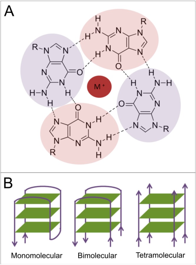
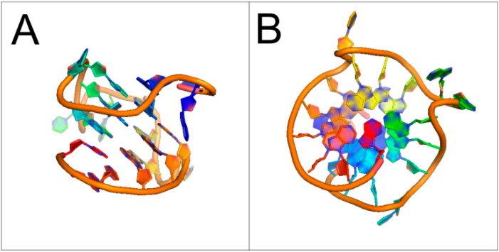
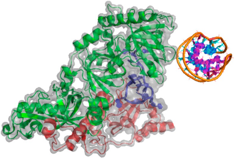
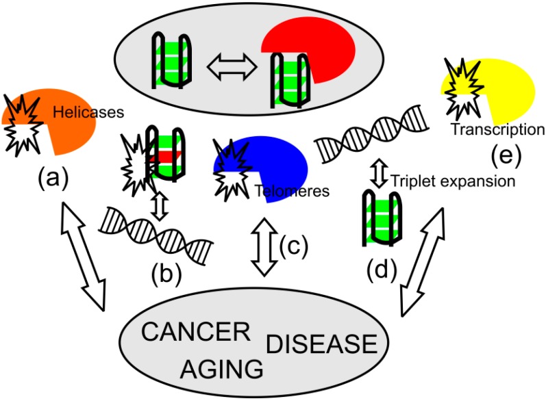
Four-stranded DNA structures were structurally characterized in vitro by NMR, X-ray and Circular Dichroism spectroscopy in detail. Among the different types of quadruplexes (i-Motifs, minor groove quadruplexes, G-quadruplexes, etc.), the best described are G-quadruplexes which are featured by Hoogsteen base-paring. Sequences with the potential to form quadruplexes are widely present in genome of all organisms. They are found often in repetitive sequences such as telomeric ones, and also in promoter regions and 5' non-coding sequences. Recently, many proteins with binding affinity to G-quadruplexes have been identified. One of the initially portrayed G-rich regions, the human telomeric sequence (TTAGGG)n, is recognized by many proteins which can modulate telomerase activity. Sequences with the potential to form G-quadruplexes are often located in promoter regions of various oncogenes. The NHE III1 region of the c-MYC promoter has been shown to interact with nucleolin protein as well as other G-quadruplex-binding proteins. A number of G-rich sequences are also present in promoter region of estrogen receptor alpha. In addition to DNA quadruplexes, RNA quadruplexes, which are critical in translational regulation, have also been predicted and observed. For example, the RNA quadruplex formation in telomere-repeat-containing RNA is involved in interaction with TRF2 (telomere repeat binding factor 2) and plays key role in telomere regulation. All these fundamental examples suggest the importance of quadruplex structures in cell processes and their understanding may provide better insight into aging and disease development.


Figures

Figure 1
Figure 1
(A) Scheme of Hoogsteen base-paring in G-quadruplex structures. The stacked tetrads of guanines (highlighted-purple, violet) are stabilized by a metal ion (M+, red) in the middle of the quadruplex; and (B) Quadruplexes can be formed within a single nucleic acid strand, from two strands (as a dimer of hairpins) or from four separate DNA or RNA strands. Green planes represent the guanine tetrads. Grey lines represent the sugar-phosphate backbone, with the arrows showing polarity of the nucleic acid chains.
Figure 2
Figure 2
Structure of G-quadruplex in the nuclease hypersensitive element (NHE) III1 region of human c-MYC promoter (PDBid: 1XAV, [16]). (A) Side view; and (B) Bottom view. Sugar-phosphate backbone is represented by the orange ribbon, with the guanine bases forming the tetrads located in the middle.
Figure 3
Figure 3
Structure of the DNA G-quadruplex of an Oxytricha nova telomeric protein-DNA complex (PDBid: 1JB7) [50]. Sugar-phosphate backbone and nucleobases in the DNA quadruplex structure is depicted by the orange ribbon and purple/cyan cartoons, respectively. The α- and β-subunits of the G-quadruplex-binding protein are represented by the green and red cartoons, respectively. A single-stranded DNA is represented by the blue cartoon. Grey color highlights the electron cloud of the protein-DNA complex.
Figure 4
Figure 4
Scheme illustrating suggested roles of quadruplexes. Quadruplex formation and recognition have various functions in biological processes and their dysregulation may be associated with human disorders, as seen from mutations in quadruplex-recognition proteins, quadruplex-resolving helicases (a) and quadruplex-forming sequences (b); as well as changes in the binding affinity and stability of telomere complexes (c); and generation of new quadruplex-forming motifs via triplet expansions (d); and transcriptional alteration (e). Arrows show connection of changes associated with quadruplex formation and recognition and influence of these changes to aging and diseases progression.

References

    1. Watson J.D., Crick F.H. Molecular structure of nucleic acids; a structure for deoxyribose nucleic acid. Nature. 1953;171:737–738. - PubMed
    1. Palecek E. Local supercoil-stabilized DNA structures. Crit. Rev. Biochem. Mol. Biol. 1991;26:151–226. - PubMed
    1. Cer R.Z., Bruce K.H., Donohue D.E., Temiz N.A., Mudunuri U.S., Yi M., Volfovsky N., Bacolla A., Luke B.T., Collins J.R., et al. Searching for non-B DNA-forming motifs using nBMST (non-B DNA motif search tool) Curr. Protoc. Hum. Genet. 2012;18:11–22. - PMC - PubMed
    1. Patel P.K., Koti A.S., Hosur R.V. NMR studies on truncated sequences of human telomeric DNA: Observation of a novel A-tetrad. Nucleic Acids Res. 1999;27:3836–3843. doi: 10.1093/nar/27.19.3836. - DOI - PMC - PubMed
    1. Caceres C., Wright G., Gouyette C., Parkinson G., Subirana J.A. A thymine tetrad in d(TGGGGT) quadruplexes stabilized with Tl+/Na+ ions. Nucleic Acids Res. 2004;32:1097–1102. doi: 10.1093/nar/gkh269. - DOI - PMC - PubMed

Publication types