. 2014 Nov 4;86(21):10625-32.
doi: 10.1021/ac5024588. Epub 2014 Oct 15.

Microfluidic Western blotting of low-molecular-mass proteins

Affiliations

Microfluidic Western blotting of low-molecular-mass proteins

Rachel E Gerver et al. Anal Chem. .

Abstract

We describe a microfluidic Western blot assay (μWestern) using a Tris tricine discontinuous buffer system suitable for analyses of a wide molecular mass range (6.5-116 kDa). The Tris tricine μWestern is completed in an enclosed, straight glass microfluidic channel housing a photopatterned polyacrylamide gel that incorporates a photoactive benzophenone methacrylamide monomer. Upon brief ultraviolet (UV) light exposure, the hydrogel toggles from molecular sieving for size-based separation to a covalent immobilization scaffold for in situ antibody probing. Electrophoresis controls all assay stages, affording purely electronic operation with no pumps or valves needed for fluid control. Electrophoretic introduction of antibody into and along the molecular sieving gel requires that the probe must traverse through (i) a discontinuous gel interface central to the transient isotachophoresis used to achieve high-performance separations and (ii) the full axial length of the separation gel. In-channel antibody probing of small molecular mass species is especially challenging, since the gel must effectively sieve small proteins while permitting effective probing with large-molecular-mass antibodies. To create a well-controlled gel interface, we introduce a fabrication method that relies on a hydrostatic pressure mismatch between the buffer and polymer precursor solution to eliminate the interfacial pore-size control issues that arise when a polymerizing polymer abuts a nonpolymerizing polymer solution. Combined with a new swept antibody probe plug delivery scheme, the Tris tricine μWestern blot enables 40% higher separation resolution as compared to a Tris glycine system, destacking of proteins down to 6.5 kDa, and a 100-fold better signal-to-noise ratio (SNR) for small pore gels, expanding the range of applicable biological targets.


Figures

Figure 1
Figure 1
Low-molecular-mass μWestern. Conducted in an enclosed microchannel filled with photoactive PA gel, the assay is comprised of three steps: (1) protein sizing after transient isotachophoresis, (2) immobilization of proteins on gel via UV photocapture (blotting), and (3) in situ antibody probing via electrophoresis. Optimization for low-molecular-mass species focuses on the separation and probing stages (bold labels). The applied electric potential is indicated by plus (+) and minus (−) symbols. Arrows indicate the direction of species electromigration under the conditions used.
Figure 2
Figure 2
Optimization of discontinuous buffer system for low-molecular-mass PAGE. (A) PAGE kymograph of Tris glycine (top) and Tris tricine (bottom) discontinuous buffer systems in a 12%T discontinuous gel; PAGE is operated under a fixed current of 1.5 μA for Tris tricine and 1 μA for Tris glycine, yielding a voltage ramp of ∼25–55 V/cm during each separation. (B) ITP sample stacking intensity profiles for protein ladder stack in open-channel regions for both the Tris glycine (upper) and Tris tricine (lower) systems at an initial sample loading and minimum sample width. During stacking, a 1.5 μA fixed current is applied for Tris tricine (∼12–25 V ramp) and a 0.3 μA fixed current (∼4–8 V ramp) for Tris glycine (as lower current yielded better stacking). Inset shows ITP stacking in a 4%T stacking gel for the Tris glycine system, added to reduce putative EOF-induced dispersion. (C) Inverted fluorescence micrographs and corresponding intensity profiles of sizing in the Tris glycine (top, open-channel loading, no 4%T gel) and Tris tricine (bottom) systems. In both cases, the 25 kDa ladder protein is observed at the 1.5 mm separation distance position.
Figure 3
Figure 3
A larger pore-size gradient at the open-channel/gel interface reduces unwanted size-exclusion effects during probing. (A) Inverted fluorescence micrographs show antibody probing across a gel with smaller pore sizes at the interface (left) and for a gel with a gradient to larger pore sizes at the interface (right), both with 12%T gels utilizing DHEBA cross-linker and 600 nM purified PSA sample. Gel interface is marked with black arrow; expected location of the PSA major isoform is indicated with an asterisk (*). (B) Inverted fluorescence kymographs of a 116–6.5 kDa ladder separation in an 8%T (top) and 12%T gel (bottom) with a Tris tricine discontinuous buffer. Right panel shows the ladder when the 25 kDa marker is 1.5 mm into the gel. In the 8%T gel, the small 6.5 kDa marker migrates faster than the stack and so rejoins the stack a short distance into the gel. 12%T enables destacking and separation of full 116–6.5 kDa ladder. (C) Schematic depicting fabrication protocol yielding a short larger-than-bulk to bulk pore-size gradient at the separation gel interface.
Figure 4
Figure 4
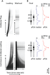
Antibody probing scheme impacts background signal. (A) Inverted fluorescence micrographs for electrophoresis of antibody probe via swept plug introduction (top) and continuous front loading (bottom). Loading images use an exposure time of 50 ms; washout images use an exposure time of 300 ms. The antibody loading concentration is 500 nM; E = 200 V/cm. (B) Inverted fluorescence micrograph showing the protein ladder and final probe results for PSA for each method. The PSA primary isoform is indicated with an asterisk (*).

Similar articles

Cited by

References

    1. Soundy P.; Harvey B. In Medical Biomethods Handbook; Walker J. M., Rapley R., Eds.; Humana Press: Totowa, NJ, 2005; pp 43–62.
    1. Allain J.-P.; Paul D.; Laurian Y.; Senn D. Members of the AIDS–Haemophilia French Study Group. Lancet 1986, 328, 1233–1236. - PubMed
    1. Shayesteh L.; Lu Y.; Kuo W.-L.; Baldocchi R.; Godfrey T.; Collins C.; Pinkel D.; Powell B.; Mills G. B.; Gray J. W. Nat. Genet. 1999, 21, 99–102. - PubMed
    1. Ghaemmaghami S.; Huh W.-K.; Bower K.; Howson R. W.; Belle A.; Dephoure N.; O’Shea E. K.; Weissman J. S. Nature 2003, 425, 737–741. - PubMed
    1. Dalmau J.; Furneaux H. M.; Gralla R. J.; Kris M. G.; Posner J. B. Ann. Neurol. 1990, 27, 544–552. - PubMed

Publication types

MeSH terms