. 2014 Nov 15;307(10):E944-53.
doi: 10.1152/ajpendo.00291.2014. Epub 2014 Sep 30.

Activation of corticotropin-releasing factor receptors in the rostral ventrolateral medulla is required for glucose-induced sympathoexcitation

Affiliations

Activation of corticotropin-releasing factor receptors in the rostral ventrolateral medulla is required for glucose-induced sympathoexcitation

Megan E Bardgett et al. Am J Physiol Endocrinol Metab. .

Abstract

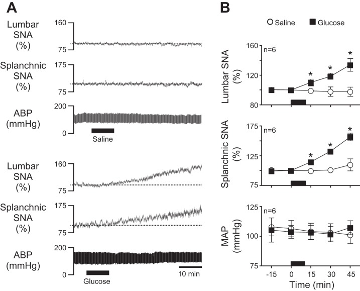
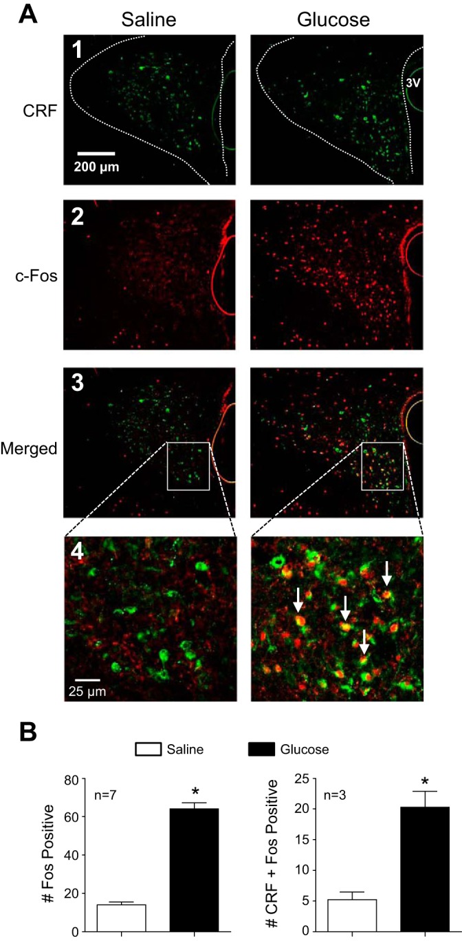
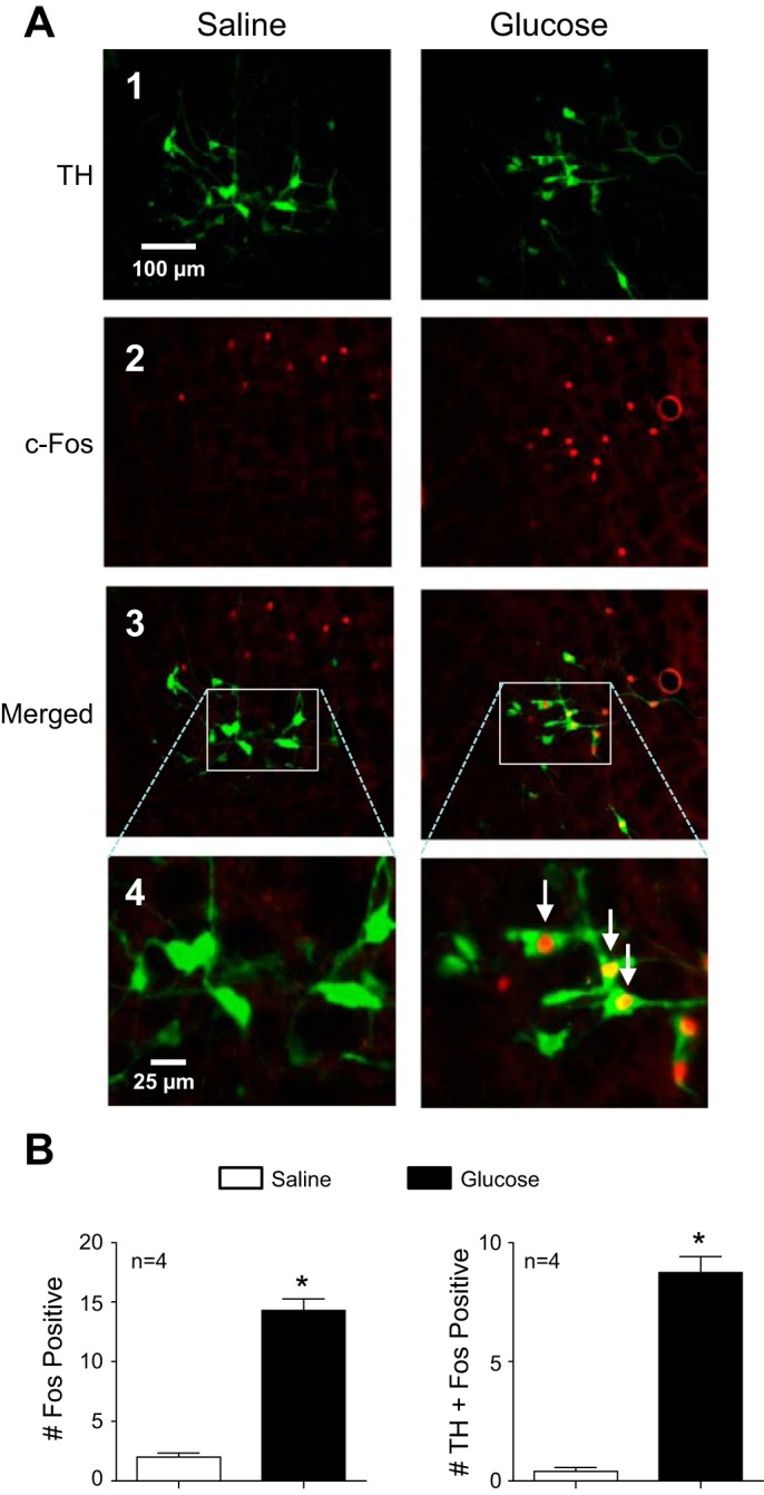
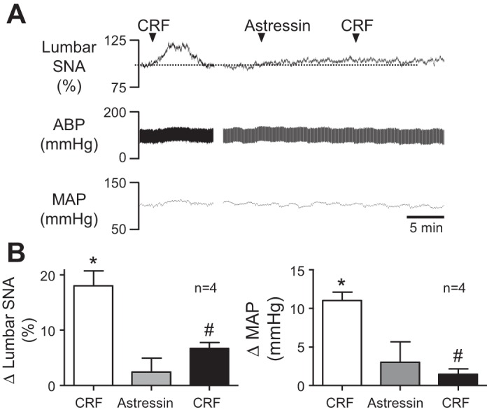
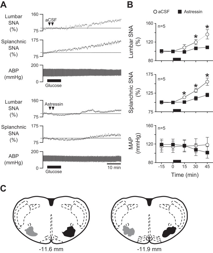
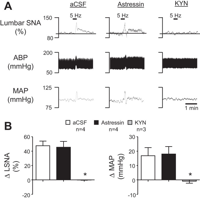
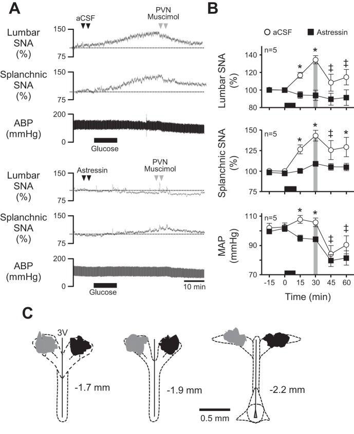
Energy expenditure is determined by metabolic rate and diet-induced thermogenesis. Normally, energy expenditure increases due to neural mechanisms that sense plasma levels of ingested nutrients/hormones and reflexively increase sympathetic nerve activity (SNA). Here, we investigated neural mechanisms of glucose-driven sympathetic activation by determining contributions of neuronal activity in the hypothalamic paraventricular nucleus (PVN) and activation of corticotropin-releasing factor (CRF) receptors in the rostral ventrolateral medulla (RVLM). Glucose was infused intravenously (150 mg/kg, 10 min) in male rats to raise plasma glucose concentration to a physiological postprandial level. In conscious rats, glucose infusion activated CRF-containing PVN neurons and TH-containing RVLM neurons, as indexed by c-Fos immunofluorescence. In α-chloralose/urethane-anesthetized rats, glucose infusion increased lumbar and splanchnic SNA, which was nearly prevented by prior RVLM injection of the CRF receptor antagonist astressin (10 pmol/50 nl). This cannot be attributed to a nonspecific effect, as sciatic afferent stimulation increased SNA and ABP equivalently in astressin- and aCSF-injected rats. Glucose-stimulated sympathoexcitation was largely reversed during inhibition of PVN neuronal activity with the GABA-A receptor agonist muscimol (100 pmol/50 nl). The effects of astressin to prevent glucose-stimulated sympathetic activation appear to be specific to interruption of PVN drive to RVLM because RVLM injection of astressin prior to glucose infusion effectively prevented SNA from rising and prevented any fall of SNA in response to acute PVN inhibition with muscimol. These findings suggest that activation of SNA, and thus energy expenditure, by glucose is initiated by activation of CRF receptors in RVLM by descending inputs from PVN.

Keywords: corticotropin releasing factor; rostral ventrolateral medulla; sympathetic nerve activity.


Figures

Fig. 1.
Fig. 1.
Acute effects of glucose infusion on lumbar sympathetic nerve activity (SNA), splanchnic SNA, and arterial blood pressure (ABP). A: representative traces of lumbar SNA, splanchnic SNA, and ABP during intravenous (iv) infusion (black bars) of saline (top) or glucose (bottom) for 10 min. Note that glucose but not saline promptly increased lumbar and splanchnic SNA. As expected, neither glucose nor saline altered ABP. B: corresponding summary data (n = 6) showing that glucose-stimulated increases of SNA reached statistical significance within 15 min and continued increasing thereafter. The period of saline/glucose infusion is indicated in each graph by the black horizontal bar along the x-axis. *P < 0.05 vs. saline. MAP, mean arterial pressure.
Fig. 2.
Fig. 2.
Effect of glucose infusion on c-Fos expression in corticotropin-releasing factor (CRF) neurons of the paraventricular nucleus (PVN). A: representative PVN sections from a saline-infused (left) and glucose-infused (right) rat showing neurons immunoreactive for CRF (image 1), c-Fos (image 2), and a merged image (image 3). In a higher-magnification merged image (image 4), numerous double-labeled neurons (arrows) can be seen only in tissue from the glucose-infused rat. B: summary data from saline- (open bar) and glucose-infused (black bar) rats (n = 3–7) revealed a significantly greater average no. of c-Fos-positive nuclei per PVN section in the glucose-infused group compared with saline-infused controls (left). The average no. of CRF-containing neurons that were colabeled with c-Fos immunoreactivity was also increased significantly by glucose infusion (right). *P < 0.05 vs. saline.
Fig. 3.
Fig. 3.
Effect of glucose infusion on c-Fos expression in tyrosine hydroxylase (TH) neurons of rostral ventrolateral medulla (RVLM). A: representative RVLM sections from a saline-infused (left) and glucose-infused (right) rat showing neurons immunoreactive for TH (image 1), c-Fos (image 2), and a merged image (image 3). In a higher-magnification merged image (image 4), numerous double-labeled neurons (arrows) can be seen only in tissue from the glucose-infused rat. B: summary data from saline- (open bar) and glucose-infused (black bar) rats (n = 4) revealed a significantly greater average no. of c-Fos positive nuclei per RVLM section in the glucose-infused group compared with saline infused controls (left). The average no. of TH-containing neurons that were colabeled with c-Fos immunoreactivity was also increased significantly by glucose infusion (right). *P < 0.05 vs. saline.
Fig. 4.
Fig. 4.
Effect of RVLM CRF receptor blockade on CRF-induced sympathoexcitation. A: representative responses of lumbar SNA, ABP, and MAP to RVLM injection of CRF before (left) and after (right) RVLM injection of the CRF antagonist astressin. Note that astressin nearly prevented sympathoexcitatory and pressor responses to CRF. B: corresponding summary data (n = 4) showing that RVLM CRF (25 pmol/50 nl) significantly (P < 0.05) increased lumbar SNA (left) and MAP (right) relative to baseline. Whereas astressin (10 pmol/50 nl) alone had no effect on either recorded variable, it significantly (P < 0.05) attenuated responses to subsequent injection of CRF. *P < 0.05 vs. baseline; #P < 0.05 vs. CRF before astressin.
Fig. 5.
Fig. 5.
Effect of RVLM CRF receptor blockade on glucose-induced sympathoexcitation. A: representative examples of lumbar SNA, splanchnic SNA, and ABP during a 10-min iv glucose infusion in which artificial cerebrospinal fluid (aCSF; top) or astressin (bottom) was bilaterally microinjected into the RVLM 5 min before the start of iv glucose infusion. B: corresponding summary data showing that RVLM pretreatment with saline or astressin had no effect on baseline SNA or MAP. In contrast, astressin significantly blunted only glucose-stimulated increases of SNA. C: schematic drawings of 2 rostrocaudal coronal planes through the RVLM showing the location of aCSF (gray; left) and astressin (black; right) injection sites. Note that injection sites were similarly placed bilaterally but are shown unilaterally for clarity. The period of glucose infusion is indicated in each part by the black horizontal bar along the x-axis. *P < 0.05 vs. aCSF.
Fig. 6.
Fig. 6.
Effect of CRF receptor blockade on the sympathoexcitatory response to sciatic afferent stimulation. A: representative examples of lumbar SNA, ABP, and MAP following aCSF, astressin, or KYN microinjection into the RVLM in response to electrical stimulation of the sciatic nerve at 5 Hz. B: summary data of the change in lumbar SNA and MAP following sciatic nerve stimulation. There was no significant difference between aCSF- and astressin-treated rats, although KYN significantly blunted the response. *P < 0.05 vs. aCSF.
Fig. 7.
Fig. 7.
Effect of RVLM CRF receptor blockade on PVN-driven sympathetic activation by glucose. A: representative responses of lumbar SNA, splanchnic SNA, and ABP to bilateral RVLM injection of aCSF (top) or astressin (bottom) prior to iv glucose infusion and PVN injection of muscimol. B: corresponding summary data showing that PVN inhibition with muscimol restored glucose-stimulated lumbar SNA and splanchnic SNA while also reducing MAP in rats pretreated with aCSF in the RVLM. In contrast, PVN inhibition had no effect when glucose responses were prevented by prior RVLM injection of astressin. C: schematic drawings of 3 rostrocaudal coronal planes through the PVN showing the location of injected muscimol in rats infused with saline (gray; left) and glucose (black; right). The period of glucose infusion is indicated in each part by the black horizontal bar along the x-axis. *P < 0.05 vs. saline; ‡P < 0.05 vs. 30 min.

References

    1. Bardgett ME, Chen QH, Guo Q, Calderon AS, Andrade MA, Toney GM. Coping with dehydration: sympathetic activation and regulation of glutamatergic transmission in the hypothalamic PVN. Am J Physiol Regul Integr Comp Physiol 306: R804–R813, 2014. - PMC - PubMed
    1. Bardgett ME, Holbein WW, Herrera-Rosales M, Toney GM. Ang II-salt hypertension depends on neuronal activity in the hypothalamic paraventricular nucleus but not on local actions of tumor necrosis factor-alpha. Hypertension 63: 527–534, 2014. - PMC - PubMed
    1. Bardgett ME, McCarthy JJ, Stocker SD. Glutamatergic receptor activation in the rostral ventrolateral medulla mediates the sympathoexcitatory response to hyperinsulinemia. Hypertension 55: 284–290, 2010. - PMC - PubMed
    1. Beckstead MJ, Gantz SC, Ford CP, Stenzel-Poore MP, Phillips PE, Mark GP, Williams JT. CRF enhancement of GIRK channel-mediated transmission in dopamine neurons. Neuropsychopharmacology 34: 1926–1935, 2009. - PMC - PubMed
    1. Cassaglia PA, Hermes SM, Aicher SA, Brooks VL. Insulin acts in the arcuate nucleus to increase lumbar sympathetic nerve activity and baroreflex function in rats. J Physiol 589: 1643–1662, 2011. - PMC - PubMed

Publication types

MeSH terms

LinkOut - more resources