. 2015 Mar;8(2):429-40.
doi: 10.1038/mi.2014.81. Epub 2014 Oct 1.

MAIT cells are licensed through granzyme exchange to kill bacterially sensitized targets

Affiliations

MAIT cells are licensed through granzyme exchange to kill bacterially sensitized targets

A Kurioka et al. Mucosal Immunol. 2015 Mar.

Abstract

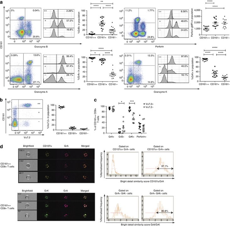
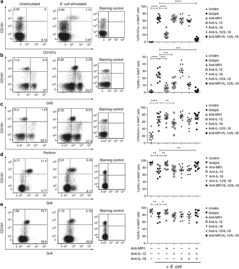
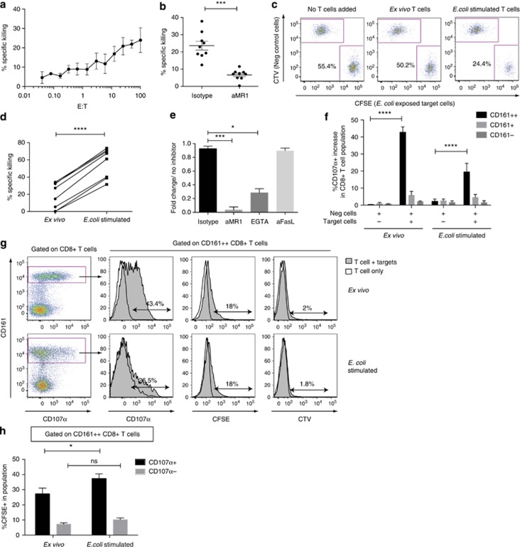
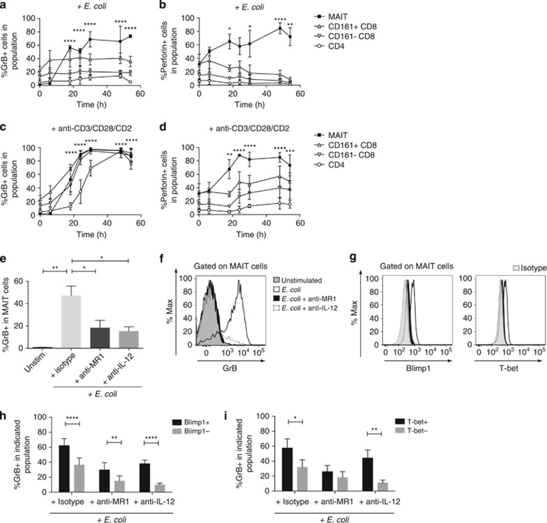
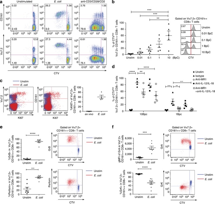
Mucosal-associated invariant T (MAIT) cells are an innate-like T-cell population restricted by the non-polymorphic, major histocompatibility complex class I-related protein 1, MR1. MAIT cells are activated by a broad range of bacteria through detection of riboflavin metabolites bound by MR1, but their direct cytolytic capacity upon recognition of cognate target cells remains unclear. We show that resting human MAIT cells are uniquely characterized by a lack of granzyme (Gr) B and low perforin expression, key granule proteins required for efficient cytotoxic activity, but high levels of expression of GrA and GrK. Bacterial activation of MAIT cells rapidly induced GrB and perforin, licensing these cells to kill their cognate target cells. Using a novel flow cytometry-based killing assay, we show that licensed MAIT cells, but not ex vivo MAIT cells from the same donors, can efficiently kill Escherichia coli-exposed B-cell lines in an MR1- and degranulation-dependent manner. Finally, we show that MAIT cells are highly proliferative in response to antigenic and cytokine stimulation, maintaining high expression of GrB, perforin, and GrA, but reduced expression of GrK following antigenic proliferation. The tightly regulated cytolytic capacity of MAIT cells may have an important role in the control of intracellular bacterial infections, such as Mycobacterium tuberculosis.


Figures

Figure 1
Figure 1
Resting blood-derived human mucosal-associated invariant T ( MAIT) cells have a unique cytotoxic profile. (a) Expression of granzyme (Gr) B, perforin, GrA, and GrK in peripheral blood CD8+ T cells from healthy donors according to CD161 expression levels. For each marker, representative staining gated on CD8+ T cells (left), representative staining gated on CD161++, CD161+, and CD161 CD8+ T cells (middle), and cumulative data for 12–15 healthy individuals (right) are shown. Results shown as mean±s.e.m., analyzed by repeated-measures one-way analysis of variance (ANOVA), with Bonferroni's multiple comparisons test. *P<0.05, **P<0.01, ***P<0.001, ****P<0.0001. (b) CD161++CD8+ T cells are predominantly Vα7.2+, compared with CD161+ and CD161 CD8+ T cells. Representative staining of Vα7.2 T-cell receptor (TCR) on CD8+ T cells according to CD161 expression levels and cumulative data are shown (n=8). (c) Expression of GrB, perforin, GrA, and GrK in Vα7.2+ and Vα7.2 cells, gated within the CD161++CD8+ T-cell population. Cumulative data are shown, analyzed by paired t-test (n=8–12). (d) Representative images showing Imagestream analysis of colocalization of GrA, GrK, and CD107α expression within CD161++CD8+ T cells. Histograms of normalized cell frequency against colocalization as measured by Bright Detail Similarity scores; >2.0 is defined as colocalized (see Supplementary Methods).
Figure 2
Figure 2
Bacterial stimulation leads to degranulation and changes in cytotoxic profile of mucosal-associated invariant T (MAIT) cells. Enriched CD8+ T cells were cocultured with THP1 cells exposed to E. coli, in the presence or absence of blocking antibodies against MR1, interleukin (IL)-12, IL-18 or isotype controls, and assayed for the expression of (a) CD107α, (b) granzyme (Gr) B, (c) perforin, (d) GrK, and (e) GrA. Representative plots gated on CD8+ T cells are shown on the left panel and cumulative data on the right panel for each marker, with bars indicating mean±s.e.m. (n=7–10). Staining control shows isotype control (conjugated with matched fluorochromes) used for gating for each marker. Note that GrA and GrK staining control is the same for each donor as both conjugated to the same flurochrome, and CD107α staining uses the unstimulated control for gating. Results are analyzed by repeated-measures one-way analysis of variance (ANOVA), with Bonferroni's multiple comparisons test. *P<0.05, **P<0.01, ***P<0.001, ****P<0.0001.
Figure 3
Figure 3
E.coli-stimulated mucosal-associated invariant T (MAIT) cells can efficiently kill target cells in an MR1- and degranulation-dependent manner. B-cell lines (BCLs) were either incubated with paraformaldehyde (PFA)-fixed E. coli or sterility control and stained with carboxyfluorescein succinimidyl ester (CFSE) and CellTrace Violet (CTV) dyes, respectively, and cocultured with enriched CD8+ T cells. (a) Percentage of specific killing of target BCLs by ex vivo MAIT cells at various E:T ratios. Mean±s.e.m. of duplicate results of three independent experiments shown (n=10). (b) Percentage of specific killing of target cells by ex vivo MAIT cells with and without an anti-MR1-blocking antibody at E:T=50:1. (n=9). (c) Example plots showing the frequency of live E. coli-exposed BCLs and negative control BCLs without effector cells (left), with ex vivo MAIT cells (middle), or with MAIT cells stimulated with E. coli for 6 days (right), added at E:T=10:1. (d) Percentage of specific killing of target cells by ex vivo and E. coli-stimulated MAIT cells from the same donors at E:T=10:1. Mean±s.e.m. of duplicate results of three independent experiments are shown. (n=8). (e) Inhibition of killing at E:T=10:1 by E. coli-stimulated MAIT cells in presence of anti-MR1 antibody, ethylene glycol tetra-acetic acid (EGTA), or anti-FasL antibody, compared with killing with no inhibitor. Repeated-measures one-way analysis of variance (ANOVA), with Bonferroni's multiple comparisons test. (n=5) (f) Cumulative data showing increase in %CD107α+ cells in indicated CD8+ T-cell populations compared with wells with T cells only. Analyzed by two-way ANOVA, comparing increase in CD107α+ expressing cells when target cells are added, within each CD8+ T-cell population, with Bonferroni's multiple comparisons test. CFSE+ E. coli-exposed BCL=Target cells, CTV+ negative control BCL=Neg cells. (n=8). (g) Representative plots of ex vivo and E. coli-stimulated CD8 T cells from the killing assays. Histograms showing expression of CD107α, and CFSE and CTV acquired through trogocytosis in CD161++CD8+ T cells. (h) Trogocytosis of CFSE from E. coli-exposed BCL membranes onto degranulating CD161++CD8+ T cells (n=8). Analyzed by two-way ANOVA, comparing wells with ex vivo and E. coli-stimulated cells, with Bonferroni's multiple comparisons test. NS, nonsignificant. *P<0.05, ***P<0.001, ****P<0.0001.
Figure 4
Figure 4
Granzyme (Gr) B and perforin upregulation in E. coli-stimulated mucosal-associated invariant T (MAIT) cells is rapid and associated with Blimp1 and T-bet expression. (ad) Peripheral blood mononuclear cells (PBMCs) were stimulated with (a, b) paraformaldehyde (PFA)-fixed E. coli, or (c, d) anti-CD3/CD28/CD2 coated beads, and stained for GrB and perforin at the indicated time points (n=3). Asterisks (*) indicate significant increases in effector molecules in MAIT cells compared with the unstimulated controls, by a two-way analysis of variance (ANOVA), with Bonferroni's multiple comparisons test. (ei) PBMCs were stimulated with PFA-fixed E. coli for 24 h with and without blocking antibodies to MR1 and interleukin (IL)-12, and stained for GrB, T-bet, and Blimp1 (n=7). (e) Cumulative data for frequency of GrB expressing MAIT cells and (f) representative example of GrB expression in MAIT cells after E. coli stimulation, ±blocking antibodies to MR1 or IL-12. One-way ANOVA, Bonferroni's multiple comparisons test. (g) Representative T-bet and Blimp1 expression in MAIT cells unstimulated or stimulated with PFA-fixed E. coli±blocking antibodies to MR1 or IL-12. For figure legend, see panel f. (h) Frequency of GrB-expressing cells in T-bet+ or T-bet MAIT cells. (i) Frequency of GrB-expressing cells in Blimp1+ or Blimp1 MAIT cells. Results analyzed by two-way ANOVA with Bonferroni's multiple comparisons test. Bars indicate mean±s.e.m. *P<0.05, **P<0.01, ***P<0.001, ****P<0.0001.
Figure 5
Figure 5
Mucosal-associated invariant T (MAIT) cells are readily proliferative in response to E. coli and maintain a highly cytotoxic profile. Peripheral blood mononuclear cells (PBMCs) from healthy donors were CellTrace Violet (CTV)-labeled and cultured with paraformaldehyde (PFA)-fixed E. coli for 6 days. BpC=bacteria per cell ratio. (a) Representative plot showing dilution of CTV against CD161 expression (upper panel) and Vα7.2 expression (lower panel) at 10 BpC or with anti-CD3/CD28/CD2 beads, gated on CD8+ T cells. (b) Cumulative data (left) and representative flow cytometry histograms are shown for each BpC. One-way analysis of variance (ANOVA), with Bonferroni's multiple comparisons test. (c) Representative plot (left) and cumulative data (right) showing Ki67 staining in CD8+ T cells after E. coli-stimulation at 10 BpC (n=4). (d) Proliferation of Vα7.2+CD161++CD8+ T cells in response to E. coli with anti-MR1, interleukin (IL)-12, and IL-18-blocking antibodies at the indicated BpCs. Results pooled from two independent experiments and analyzed by two-way ANOVA, with Bonferroni's multiple comparisons test. (e) GrB and perforin, and (f) GrA and GrK expression in CTV-labeled Vα7.2+CD161++CD8+ T cells following E. coli stimulation for 6 days. Cumulative data (left) and representative staining is shown (right). Gating for granzymes and perforin based on isotype controls. Results analyzed by paired t-test (n=8). Bars indicate mean±s.e.m. *P<0.05, **P<0.01, ***P<0.001, ****P<0.0001.

References

    1. Treiner E., et al. Selection of evolutionarily conserved mucosal-associated invariant T cells by MR1. Nature. 2003;422:164–169. - PubMed
    1. Le Bourhis L., et al. Antimicrobial activity of mucosal-associated invariant T cells. Nat. Immunol. 2010;11:701–708. - PubMed
    1. Gold M.C., et al. Human mucosal associated invariant T cells detect bacterially infected cells. PLoS Biol. 2010;8:e1000407. - PMC - PubMed
    1. Chua W.J., et al. Polyclonal mucosal-associated invariant T cells have unique innate functions in bacterial infection. Infect. Immun. 2012;80:3256–3267. - PMC - PubMed
    1. Georgel P., Radosavljevic M., Macquin C., Bahram S. The non-conventional MHC class I MR1 molecule controls infection by Klebsiella pneumoniae in mice. Mol. Immunol. 2011;48:769–775. - PubMed

Publication types

MeSH terms