

Cellular proliferation in the nasal septal cartilage of juvenile minipigs
- PMID: 25269781
- PMCID: PMC4262346
- DOI: 10.1111/joa.12237
Cellular proliferation in the nasal septal cartilage of juvenile minipigs
Abstract
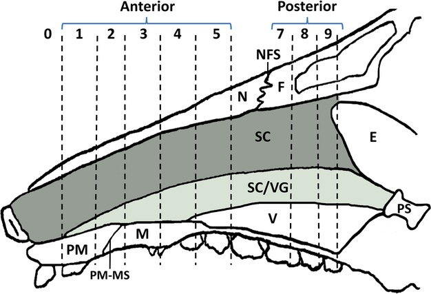
The growth of the nasal septal cartilage is believed to be a driving force of midfacial growth. Cellular proliferation is an important contributor to growth of the cartilage, but this factor has been rarely investigated. The current study was undertaken to assess the proliferation and cellular density in the septal cartilage of fast-growing juvenile minipigs. Six minipigs averaging 4.4 ± 1 months old were injected with 5'-bromo-2'-deoxyuridine (BrdU), a thymidine analog, 24 h before death. The septal cartilage was sectioned in the coronal plane and reacted for BrdU. The proliferative index (number of BrdU-positive chondrocytes/total number of chondrocytes) and cellular density (number of cells mm(-2) ) of various locations of the septum were measured and compared in order to determine overall proliferation rate and whether regional variations in proliferative activity and cellular density are present. To provide a time perspective to the problem of midfacial growth, the lengths of the nasal bone and the palate were measured in a collection of 61 dry skulls of minipigs aged 1-8 months. Results showed that the septal chondrocytes were proliferating at a surprisingly high rate (~21%). The proliferative index was higher in the ventral and middle compared with the dorsal locations, and in the central cartilage compared with the perichondrium. No difference in proliferative index was found between the anterior and posterior parts of the septum. Cellular density was higher in the perichondrium than in the central cartilage. Within the central cartilage there was a trend for higher cellular density anteriorly. In conclusion, the rapidly growing midface of juvenile minipigs is associated with a high rate of septal proliferation, especially in the ventral half of the cartilage.
Keywords: 5′-bromo-2′-deoxyuridine; cellular proliferation; midfacial growth; minipigs; nasal cartilage.
© 2014 Anatomical Society.
Figures


References
-
- Al Dayeh AA, Rafferty KL, Egbert M, et al. Real-time monitoring of the growth of the nasal septal cartilage and the nasofrontal suture. Am J Orthod Dentofac Orthop. 2013;143:773–783. - PubMed
-
- Babler WJ, Persing JA, Persson KM, et al. Skull growth after coronal suturectomy, periostectomy, and dural transection. J Neurosurg. 1982;56:529–535. - PubMed
-
- Carnevale GG, Tatum SA. Chondrocyte volume distribution in porcine septal cartilage: an initial stereoscopic evaluation. Otolaryngol Head Neck Surg. 1996;115:365–369. - PubMed
-
- Carnevale GG, Tatum SA, Malmgren LT. 2000. Stereological assessment of porcine septal chondroctye distribution. Unpublished presentation from Annual Fall Meeting of the American Academy of Facial Plastic and Reconstructive Surgery, Sept. 21–23.
Publication types
MeSH terms
Grants and funding
LinkOut - more resources
Full Text Sources
Other Literature Sources
