Review
. 2015 Jan;235(2):185-95.
doi: 10.1002/path.4454.

Molecular pathology of emerging coronavirus infections

Affiliations
Review

Molecular pathology of emerging coronavirus infections

Lisa E Gralinski et al. J Pathol. 2015 Jan.

Abstract

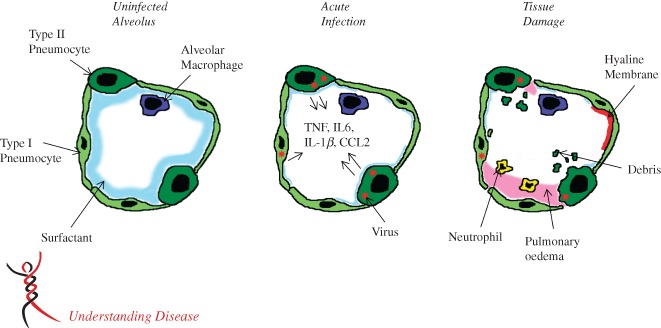
Respiratory viruses can cause a wide spectrum of pulmonary diseases, ranging from mild, upper respiratory tract infections to severe and life-threatening lower respiratory tract infections, including the development of acute lung injury (ALI) and acute respiratory distress syndrome (ARDS). Viral clearance and subsequent recovery from infection require activation of an effective host immune response; however, many immune effector cells may also cause injury to host tissues. Severe acute respiratory syndrome (SARS) coronavirus and Middle East respiratory syndrome (MERS) coronavirus cause severe infection of the lower respiratory tract, with 10% and 35% overall mortality rates, respectively; however, >50% mortality rates are seen in the aged and immunosuppressed populations. While these viruses are susceptible to interferon treatment in vitro, they both encode numerous genes that allow for successful evasion of the host immune system until after high virus titres have been achieved. In this review, we discuss the importance of the innate immune response and the development of lung pathology following human coronavirus infection.

Keywords: ARDS; MERS-CoV; SARS-CoV; acute lung injury; acute respiratory distress syndrome; coronavirus; type II pneumocytes.


Figures

Figure 1
Figure 1
MA‐SARS lung immunopathology. (A) Mock‐infected lung stained with haematoxylin and eosin. (B) Large airway of a C57BL/6 J (B6) mouse, 7 days post‐infection, with 105 plaque‐forming units (PFU) MA‐SARS, shows denudation of the epithelial cells. (C, D) Immunohistochemical staining of the SARS‐CoV N protein at 2 days post‐infection shows staining consistent with infection of airway epithelial cells and type II pneumocytes, respectively. (E) MSB staining highlights fibrin in the parenchyma of the lung (red staining) in B6 mice, 7 days post‐infection with 105 PFU MA‐SARS. (F) Perivascular cuffing in a B6 mouse, 4 days post‐infection with 105 PFU MA‐SARS. (G) Hyaline membranes in the parenchyma of the lung of a B6 mouse, 7 days post‐infection with 105 PFU MA‐SARS. (H) Inflammation in the lung of a B6 mouse, 7 days post‐infection with 104 PFU MA‐SARS. (I) Haemorrhage in the lung of a Serpine1 mouse, 7 days post‐infection with 104 PFU MA‐SARS.
Figure 2
Figure 2
Model of an infected alveolus in the lung. Type I and type II pneumocytes make up the alveolar walls and resident alveolar macrophages and pulmonary surfactant exist in the airspace (A). In the acute phase of SARS‐CoV infection (B), type I and type II pneumocytes are infected and secrete inflammatory cytokines, while surfactant levels decrease. During the late stage/tissue damage portion of viral infection, viral titres decrease, while airway debris, pulmonary oedema and hyaline membrane formation all impede respiration (C).

References

    1. Johnson ER, Matthay MA. Acute lung injury: epidemiology, pathogenesis, and treatment. J Aerosol Med Pulm Drug Deliv 2010; 23: 243–252. - PMC - PubMed
    1. Reynolds HN, McCunn M, Borg U, et al. Acute respiratory distress syndrome: estimated incidence and mortality rate in a 5 million‐person population base. Crit Care 1998; 2: 29–34. - PMC - PubMed
    1. Cabeca TK, Granato C, Bellei N. Epidemiological and clinical features of human coronavirus infections among different subsets of patients. Influenza Other Respir Viruses 2013; 7: 1040–1047. - PMC - PubMed
    1. van der Hoek L, Pyrc K, Jebbink MF, et al. Identification of a new human coronavirus. Nat Med 2004; 10: 368–373. - PMC - PubMed
    1. Woo PC, Lau SK, Chu CM, et al. Characterization and complete genome sequence of a novel coronavirus, coronavirus HKU1, from patients with pneumonia. J Virol 2005; 79: 884–895. - PMC - PubMed

Publication types

MeSH terms

Substances