. 2015 May;26(5):1053-70.
doi: 10.1681/ASN.2014030233. Epub 2014 Sep 30.

Deficiency of fibroblast growth factor-inducible 14 (Fn14) preserves the filtration barrier and ameliorates lupus nephritis

Affiliations

Deficiency of fibroblast growth factor-inducible 14 (Fn14) preserves the filtration barrier and ameliorates lupus nephritis

Yumin Xia et al. J Am Soc Nephrol. 2015 May.

Abstract

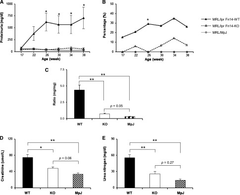
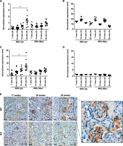
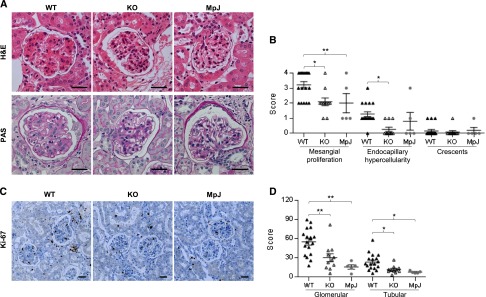
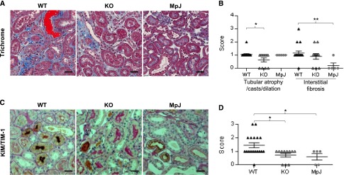
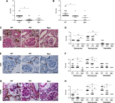
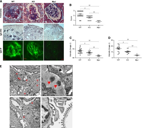
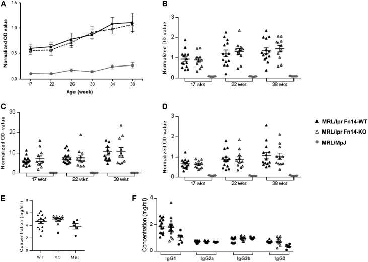
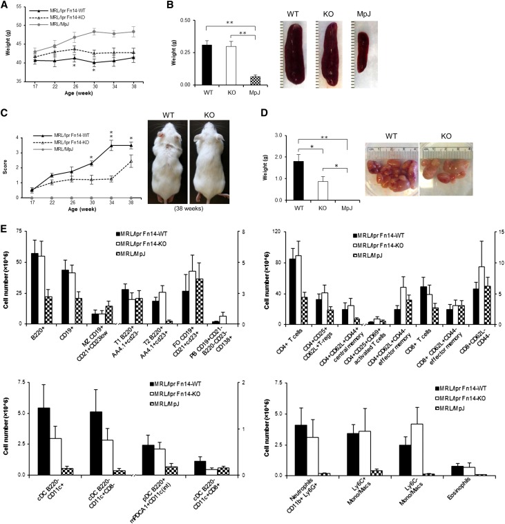
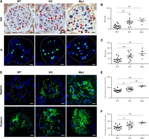
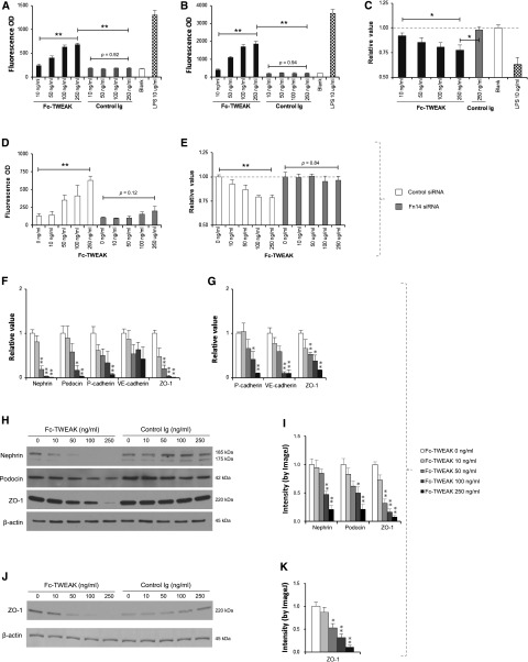
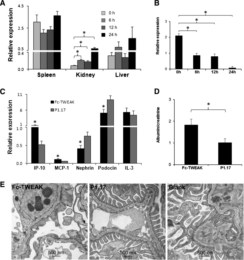
TNF ligand superfamily member 12, also known as TNF-related weak inducer of apoptosis (TWEAK), acts through its receptor, fibroblast growth factor-inducible 14 (Fn14), to mediate several key pathologic processes involved in tissue injury relating to lupus nephritis. To explore the potential for renal protection in lupus nephritis by targeting this pathway, we introduced the Fn14 null allele into the MRL-lpr/lpr lupus mouse strain. At 26-38 weeks of age, female Fn14-knockout MRL-lpr/lpr mice had significantly lower levels of proteinuria compared with female wild-type MRL-lpr/lpr mice. Furthermore, Fn14-knockout mice had significantly improved renal histopathology accompanied by attenuated glomerular and tubulointerstitial inflammation. There was a significant reduction in glomerular Ig deposition in Fn14-knockout mice, despite no detectable differences in either serum levels of antibodies or splenic immune cell subsets. Notably, we found that the Fn14-knockout mice displayed substantial preservation of podocytes in glomeruli and that TWEAK signaling directly damaged barrier function and increased filtration through podocyte and glomerular endothelial cell monolayers. Our results show that deficiency of the Fn14 receptor significantly improves renal disease in a spontaneous lupus nephritis model through prevention of the direct injurious effects of TWEAK on the filtration barrier and/or modulation of cytokine production by resident kidney cells. Thus, blocking the TWEAK/Fn14 axis may be a novel therapeutic intervention in immune-mediated proliferative GN.

Keywords: clinical immunology; cytokines; lupus nephritis.


Figures

Figure 1.
Figure 1.
Proteinuria is attenuated in MRL/lpr Fn14-KO mice. (A) Beginning at 26 weeks until the mice were euthanized at 38 weeks, there was significantly decreased proteinuria in MRL/lpr Fn14-KO compared with MRL/lpr Fn14-WT mice. The MRL/lpr Fn14-KO strain also displayed a lower percentage of mice with (B) severe proteinuria (≥300 mg/dl at 26 weeks by chi-squared test) and (C) decreased urine albumin-to-creatinine ratio (at 38 weeks). Serum levels of (D) creatinine and (E) BUN were higher in MRL/lpr Fn14-WT compared with Fn14-KO mice. The numbers of MRL/lpr Fn14-WT, MRL/lpr Fn14-KO, and MRL/MpJ mice were 14, 11, and 5, respectively. *P<0.05; **P<0.01.
Figure 2.
Figure 2.
Renal TWEAK/Fn14 expression increases with age in MRL/lpr mice. (A) mRNA expression level of TWEAK in kidneys from MRL/lpr Fn14-WT mice was higher at 26 than 7 weeks of age but revealed no difference between those two time points in MRL/MpJ mice (P>0.05). (B) mRNA expression level of TWEAK in spleens did not increase with age in either MRL/lpr or MRL/MpJ mice. (C and D) The expression levels of Fn14 mRNA in (C) kidneys and (D) spleens displayed similar trends as TWEAK in the two strains. In A–D, the numbers of mice are provided in parentheses after the age on the x axis. The experiments depicted in A–D were repeated two times with similar results. *P<0.05. (E) IHC staining verified Fn14 protein expression in the kidneys from MRL/lpr Fn14-WT mice at ages of 17, 30, and 38 weeks. MRL/lpr Fn14-KO mice at 38 weeks (n=5) show minimal background staining. Representative images are shown. Scale bar, 40 μm.
Figure 3.
Figure 3.
Kidney proliferative changes are attenuated in MRL/lpr Fn14-KO mice. (A) Representative kidney sections (stained with H&E or periodic acid–Schiff [PAS]) of three mouse strains are shown that display reduced glomerular damage in MRL/lpr Fn14-KO mice compared with MRL/lpr Fn14-WT mice. H&E, hematoxylin and eosin. (B) Semiquantitative scoring results on the basis of H&E and PAS staining revealed significant differences in histopathological features of LN between the MRL/lpr Fn14-WT and Fn14-KO mice. H&E, hematoxylin and eosin. (C) Representative images of Ki-67 staining are shown. (D) Ki-67–stained sections were scored and exhibited fewer Ki-67–positive cells in both glomerular and tubular areas in kidneys of Fn14-KO mice compared with the MRL/lpr Fn14-WT strain. There were no differences between the MRL/lpr Fn14-KO mice and MRL/MpJ mice (P>0.05). The numbers of MRL/lpr Fn14-WT, MRL/lpr Fn14-KO, and MRL/MpJ mice (all at 38 weeks of age) were 14, 11, and 5, respectively. Scale bar, 25 μm. *P<0.05; **P<0.01.
Figure 4.
Figure 4.
Kidney tubulointerestitial changes are decreased in MRL/lpr Fn14-KO mice. (A) By trichrome staining, tubular casts (red) and interstitial fibrosis (blue) could be seen in MRL/lpr Fn14-WT mice but were relatively rare in the MRL/MpJ mice. (B) Semiquantitative scoring results on the basis of trichrome staining revealed significant difference in tubular injury but not in interstitial fibrosis between the MRL/lpr Fn14-WT and Fn14-KO mice. (C) Representative images of KIM-1/TIM-1 staining are shown, displaying diffuse staining in proximal tubules in kidneys of MRL/lpr Fn14-WT mice, patchy staining in Fn14-KO mice, and slight staining in MRL/MpJ mice. (D) Semiquantitative scoring showed reduced KIM-1/TIM-1 staining in Fn14-KO mice compared with MRL/lpr Fn14-WT mice, but it was not different from MRL/MpJ mice (P>0.05). The numbers of MRL/lpr Fn14-WT, MRL/lpr Fn14-KO, and MRL/MpJ mice (all at 38 weeks of age) were 14, 11, and 5, respectively. Scale bar, 25 μm. *P<0.05; **P<0.01.
Figure 5.
Figure 5.
Kidney cellular infiltration is attenuated in MRL/lpr Fn14-KO mice. (A and B) Semiquantitative scoring results on the basis of H&E and periodic acid–Schiff (PAS) staining revealed significant reduction of both (A) cortical (nonperivascular) and (B) perivascular inflammation in MRL/lpr Fn14-KO mice compared with MRL/lpr Fn14-WT mice. H&E, hematoxylin and eosin. (C) Representative images of B220 staining are shown, displaying intense infiltration in the perivascular areas of kidneys from MRL/lpr mice but no infiltration in MRL/MpJ mice, which was also confirmed by (D) semiquantitative scoring. (E) Representative images of CD3 staining are shown, reflecting (F) higher scores of glomerular and perivascular infiltration in MRL/lpr mice compared with MRL/MpJ mice. There were no differences in both B220 and CD3 staining between the MRL/lpr Fn14-WT and Fn14-KO mice (P>0.05). (G) Representative images of F4/80 staining are shown. (H) Sections of F4/80 staining were scored separately in the glomerular, perivascular, and interstitial areas, showing significant reduction in MRL/lpr Fn14-KO mice in all of these regions compared with MRL/lpr Fn14-WT mice. The MRL/lpr Fn14-KO mice differed from MRL/MpJ mice in F4/80 infiltration in the perivascular but not glomerular or interstitial areas. The numbers of MRL/lpr Fn14-WT, MRL/lpr Fn14-KO, and MRL/MpJ mice (all at 38 weeks of age) were 14, 11, and 5, respectively. Scale bar, 25 μm. *P<0.05; **P<0.01.
Figure 6.
Figure 6.
Glomerular IC deposition is reduced in MRL/lpr Fn14-KO mice. (A) Representative images of mouse kidneys are shown for (top panel) trichrome staining, (middle panel) IgG staining by IHC, and (bottom panel) IF. In trichrome staining, white arrows indicate IC, and black arrows indicate red blood cells. (B) Slides stained by trichrome or periodic acid–Schiff (images not shown) were used for scoring immune deposits, with higher scores in the MRL/lpr Fn14-WT than the Fn14-KO strain. (C and D) IgG deposition detected by (C) IHC and (D) IF was quantitated by ImageJ, showing a significant reduction in Fn14-KO mice compared with MRL/lpr Fn14-WT mice, although still higher than MRL/MpJ controls. (E) Representative TEM images showed that, compared with MRL/lpr Fn14-KO mice (n=3), electron-dense deposits (red arrows) were more prominent in the glomerular basement membrane of MRL/lpr Fn14-WT mice (n=2). Fusion of podocyte foot processes (black arrows) was also more severe in MRL/lpr Fn14-WT mice. Neither electron-dense deposit nor foot process fusion was seen in glomeruli in MRL/MpJ mice (n=3). For A–D, the numbers of mice in each of the MRL/lpr Fn14-WT, MRL/lpr Fn14-KO, and MRL/MpJ strains were 14, 11, and 5, respectively. For A–E, age = 38 weeks. *P<0.05; **P<0.01.
Figure 7.
Figure 7.
Serum antibody levels are not altered with Fn14 deficiency in MRL/lpr mice. (A) There were no significant differences in the titers of IgG anti-dsDNA antibodies between MRL/lpr Fn14-WT and Fn14-KO mice at any time point (P>0.05). (B–D) There were no differences between MRL/lpr Fn14-WT and Fn14-KO mice in (B) anti-chromatin, (C) anti-ribosomal P, and (D) anti-cardiolipin autoantibodies (P>0.05) at any of the time points. MRL/MpJ mice had significantly lower serum levels of these autoantibodies than the MRL/lpr strains at all time points. The experiments depicted in A–D were repeated two times with similar results. (E and F) There were no significant differences in the serum levels of (E) total IgG or (F) specific IgG isotypes between MRL/lpr Fn14-WT and Fn14-KO mice at 38 weeks of age (P>0.05). The numbers of MRL/lpr Fn14-WT, MRL/lpr Fn14-KO, and MRL/MpJ mice were 14, 11, and 5, respectively. dsDNA, double-stranded DNA.
Figure 8.
Figure 8.
Fn14 deficiency affects lymphadenopathy but not splenocyte weight or cellular composition in MRL/lpr mice. (A) MRL/lpr Fn14-WT and Fn14-KO mice exhibited less weight gain compared with MRL/MpJ mice between 26 and 38 weeks of age. A significant difference between MRL/lpr Fn14-WT and Fn14-KO mice was seen at 26 and 30 weeks. (B) At euthanasia (38 weeks of age), there were no differences in spleen weights between the MRL/lpr Fn14-KO and Fn14-WT mice. Representative images of spleens from mice at age of 38 weeks are shown. MRL/lpr Fn14-WT mice differed from Fn14-KO mice in (C) lymphadenopathy scores beginning at 30 weeks of age and (D) lymph node weight at 38 weeks. Representative images of (C) lymphadenopathy and (D) lymph nodes from MRL/lpr Fn14-WT and Fn14-KO mice at the age of 38 weeks are shown. No visibly enlarged lymph nodes were present in MRL/MpJ mice. For A–D, the numbers of MRL/lpr Fn14-WT, MRL/lpr Fn14-KO, and MRL/MpJ mice were 14, 11, and 5, respectively. (E) FACS analysis showed that, in four major subpopulations of spleen cells (T, B, dendritic, and myeloid cells), there were no significant differences between MRL/lpr Fn14-WT (n=8) and Fn14-KO (n=6) mice at week 38. Five MRL/MpJ mice were studied in the cytometric analysis. *P<0.05; **P<0.01.
Figure 9.
Figure 9.
Podocytes are protected by Fn14 deficiency in MRL/lpr mice. (A) Representative images of WT1 staining by IHC and IF are shown. (Upper panel) The brown nuclei indicated by red arrows represent WT1-positive cells. (Lower panel) By IF, WT1-positive nuclei (green) were overlapped by 4′,6-diamidino-2-phenylindole staining (blue). Single glomeruli are outlined by dashed lines. The sections were scored by counting the number of WT1-positive nuclei per glomerulus (10 random glomeruli per section). Compared with MRL/lpr Fn14-WT mice, the Fn14-KO mice had increased scores by both (B) IHC and (C) IF staining and were not significantly different from MRL/MpJ mice. (D–F) Representative images are shown (IF staining) for nephrin and podocin expressions (green), which were the strongest in MRL/MpJ mice followed by MRL/lpr Fn14-KO and Fn14-WT mice. The experiments depicted in D–F were repeated three times with similar results. The numbers of MRL/lpr Fn14-WT, MRL/lpr Fn14-KO, and MRL/MpJ mice (all at 38 weeks of age) were 14, 11, and 5, respectively. Scale bar, 10 μm. *P<0.05; **P<0.01.
Figure 10.
Figure 10.
Activation of the TWEAK/Fn14 pathway modulates the integrity of the renal filtration barrier in vitro. (A and B) Fc-TWEAK but not control Ig stimulation increased permeability for both (A) BSA-FITC and (B) dextran-FITC through podocyte monolayers in a dose-dependent manner (by Spearman rank correlation; P<0.01). LPS treatment was used as a positive control. (C) The viability assay for podocytes showed significant differences between the Fc-TWEAK 250 ng/ml group and the Fc-TWEAK 10 ng/ml or control Ig 250 ng/ml group. All values were normalized to the untreated blank group (as a baseline of one). (D and E) Fn14 siRNA but not control siRNA blocked the effects of Fc-TWEAK on both (D) permeability and (E) death of podocytes. (F) mRNA levels of nephrin, podocin, P-cadherin, and ZO-1 decreased with increasing concentrations of Fc-TWEAK in the culture media. (G) A similar finding was seen with P-cadherin, VE-cadherin, and ZO-1 mRNA expressions by glomerular endothelial cells. (H) Decreasing expressions of nephrin, podocin, and ZO-1 by podocytes with increasing concentration of Fc-TWEAK but not the control Ig are observed by Western blot. (I) The intensity of bands of Western blot were quantitated by ImageJ, showing significant decrease of nephrin, podocin, and ZO-1 by podocytes with Fc-TWEAK. (J) Glomerular endothelial cells expressed less ZO-1 protein after Fc-TWEAK stimulation, which was shown by (K) ImageJ quantitation of Western bot bands. Data were representative of three to five independent experiments. For F–K, comparisons are to media alone (Fc-TWEAK=0 ng/ml). *P<0.05; **P<0.01.
Figure 11.
Figure 11.
Exogenous TWEAK has direct kidney effects. (A and B) Female MRL/MpJ mice at 8 weeks of age (n=3 in each group) received a single intravenous injection of 200 µg Fc-TWEAK or the P1.17 isotype control, and the kidneys were obtained at euthanasia. Fc-TWEAK–injected mice displayed (A) a significant increase in kidney mRNA expression of IP-10 and (B) decreased expression of podocin at 6, 12, and 24 hours compared with baseline. There were no differences in IP-10 levels in the spleen or liver at any of these time points (P>0.05). (C–E) Predisease female MRL/lpr mice (8 weeks old) were injected with 200 µg Fc-TWEAK or P1.17 intravenously and euthanized 12 hours later. Fc-TWEAK–injected mice (n=4) had significantly higher renal mRNA expression of IP-10 and MCP-1 and lower nephrin and podocin expressions than P1.17-injected mice (n=4). (C) Levels of IL-3, a cytokine not regulated by TWEAK, were unchanged. Furthermore, TWEAK-injected mice exhibited (D) increased proteinuria and (E) podocyte foot process effacement. Representative TEM images are shown. Blank is a representative kidney section from a noninjected age- and sex-matched MRL/lpr mouse. In A–C, relative values of mRNA expression were multiplied by 102. *P<0.05.

References

    1. Borchers AT, Leibushor N, Naguwa SM, Cheema GS, Shoenfeld Y, Gershwin ME: Lupus nephritis: A critical review. Autoimmun Rev 12: 174–194, 2012 - PubMed
    1. Xia Y, Pawar RD, Nakouzi AS, Herlitz L, Broder A, Liu K, Goilav B, Fan M, Wang L, Li QZ, Casadevall A, Putterman C: The constant region contributes to the antigenic specificity and renal pathogenicity of murine anti-DNA antibodies. J Autoimmun 39: 398–411, 2012 - PMC - PubMed
    1. Lech M, Anders HJ: The pathogenesis of lupus nephritis. J Am Soc Nephrol 24: 1357–1366, 2013 - PMC - PubMed
    1. Burkly LC, Michaelson JS, Zheng TS: TWEAK/Fn14 pathway: An immunological switch for shaping tissue responses. Immunol Rev 244: 99–114, 2011 - PubMed
    1. Brown SA, Cheng E, Williams MS, Winkles JA: TWEAK-independent Fn14 self-association and NF-κB activation is mediated by the C-terminal region of the Fn14 cytoplasmic domain. PLoS ONE 8: e65248, 2013 - PMC - PubMed

Publication types