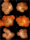
Retinal changes in visceral leishmaniasis by retinal photography
- PMID: 25270641
- PMCID: PMC4261886
- DOI: 10.1186/1471-2334-14-527
Retinal changes in visceral leishmaniasis by retinal photography
Abstract
Background: In visceral leishmaniasis (VL), retinal changes have previously been noted but not described in detail and their clinical and pathological significance are unknown. A prospective observational study was undertaken in Mymensingh, Bangladesh aiming to describe in detail visible changes in the retina in unselected patients with VL.
Methods: Patients underwent assessment of visual function, indirect and direct ophthalmoscopy and portable retinal photography. The photographs were assessed by masked observers including assessment for vessel tortuosity using a semi-automated system.
Results: 30 patients with VL were enrolled, of whom 6 (20%) had abnormalities. These included 5 with focal retinal whitening, 2 with cotton wool spots, 2 with haemorrhages, as well as increased vessel tortuosity. Visual function was preserved.
Conclusions: These changes suggest a previously unrecognized retinal vasculopathy. An inflammatory aetiology is plausible such as a subclinical retinal vasculitis, possibly with altered local microvascular autoregulation, and warrants further investigation.
Figures
References
-
- Acharyya C. Retinal haemorrhage in kala-azar. J Indian Med Assoc. 1957;28:437. - PubMed
-
- Ling WP. Ocular changes in kala-azar in Peking. Am J Ophthalmol. 1924;7:829–834. doi: 10.1016/S0002-9394(24)90891-9. - DOI
-
- De Cock KM, Rees PH, Klauss V, Kasili EG, Kager PA, Schattenkerk JK. Retinal hemorrhages in kala-azar. Am J Trop Med Hyg. 1982;31:927–930. - PubMed
-
- Mookerjee GC, Sen G, Chaudhuri MD, Chakraborty K. Acute kala-azar with haemorrhagic retinopathy. J Indian Med Assoc. 1975;65:86–88. - PubMed
Pre-publication history
-
- The pre-publication history for this paper can be accessed here:http://www.biomedcentral.com/1471-2334/14/527/prepub
Publication types
MeSH terms
Grants and funding
LinkOut - more resources
Full Text Sources
Other Literature Sources
Medical


