Review
. 2014 Nov-Dec;3(6):489-500.
doi: 10.1002/wdev.159. Epub 2014 Sep 30.

WNT signaling in bone development and homeostasis

Affiliations
Review

WNT signaling in bone development and homeostasis

Zhendong Zhong et al. Wiley Interdiscip Rev Dev Biol. 2014 Nov-Dec.

Abstract

The balance between bone formation and bone resorption controls postnatal bone homeostasis. Research over the last decade has provided a vast amount of evidence that WNT signaling plays a pivotal role in regulating this balance. Therefore, understanding how the WNT signaling pathway regulates skeletal development and homeostasis is of great value for human skeletal health and disease.


Figures

Figure 1
Figure 1. Overview of Bone Formation
During intramembranous ossification, new bone forms directly from a condensation of mesenchymal stem cells. In contrast, endochondral ossification occurs when a cartilaginous template is replaced by mineralized osteoid. In both cases, osteoblasts become encased in mineralized matrix creating osteocytes. Please refer to the text for more detail.
Figure 2
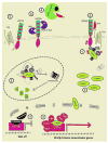
Figure 2. Canonical Wnt Signaling Pathway
In the absence of an upstream ligand, receptor Frizzled (FZD) and co-receptor LRP5/6 are inactive (1). The cytoplasmic β-catenin will be recruited into a “destruction complex” consisting of Axin, APC, casein kinase 1a (CK1) and glycogen synthase kinase 3 (GSK3) (2). This “destruction complex” facilitates the phosphorylation of β-catenin by GSK3 and subsequent ubiquitinylation (Ub) by β-TrCP, an E3 ligase, targeting β-catenin for proteasome-mediated degradation. In this context, the transcription repressor Groucho occupies T-Cell Factor/Lymphoid enhancer factor (TCF/LEF) DNA binding sites (7). This inactive state could also be caused by lack of co-receptors availability due to effectors (DKK1/WISE/SOST) binding to LRP5/6 and preventing its association with Wnt (3), or by other effectors associating with Wnts and blocking their ability to interact with the co-receptor complex (4). When a Wnt ligand engages the receptor complex (5), the C-terminus of LRP5/6 is phosphorylated, creating a binding site for Axin, resulting in inhibition of the destruction complex (6). This inhibits GSK3 activity, allowing cytoplasmic levels of β-catenin to increase (8). β-catenin subsequently translocates into the nucleus and complexes with LEF/TCF proteins and other co-factors to activate transcription of target genes (9).
Figure 3
Figure 3. Regulation of RANKL Signaling
The Receptor activator of nuclear factor κβ (RANK) is expressed on osteoclasts and upon binding from its cognate ligand, RANKL, stimulates osteoclast differentiation and activation. RANKL is expressed by cells of the osteoblast lineage, which also express another protein referred to as Osteoprotegerin (OPG). OPG binds to RANKL, prevents interaction between RANKL with RANK with the end result being inhibition of osteoclast differentiation and activation. OPG expression can also be activation at the transcriptional level via the binding of β-catenin/TCF complexes to the OPG promoter.
Figure 4
Figure 4. Low-density Lipoprotein (LDL) Receptor Family Members with Putative Roles in Bone Development
The Low-density lipoprotein related receptors are single pass Type I transmembrane proteins which share several structural motifs. These include β-propellers (each containing several individual EGF repeats) and ligand-binding repeats (which mediate interactions with apolipoproteins. The relative sizes of LRP1, LRP4, LRP5/6, and LRP8 are represented. LRP5 and LRP6 form a novel subclass of this family with the cytoplasmic tail of these proteins containing PPPS/TP motifs which are targets of phosphorylation and act to stimulate downstream signaling upon activation by WNT ligands.

References

    1. Franz-Odendaal TA. Induction and patterning of intramembranous bone. Front Biosci (Landmark Ed) 2011;16:2734–2746. - PubMed
    1. Karaplis AC. Embryonic development of bone and the molecular regulation of intramembranous and endochondral bone formation. In: Bilezikian JP, Raisz L, Rodan GA, editors. Principles of Bone Biology. 2. Vol. 1. San Diego, CA: Academic Press; 2002. pp. 33–58.
    1. Recker R. Embryology, anatomy, and microstructure of bone. In: Coe FL, Favus MJ, editors. Disorders of Bone and Mineral Metabolism. New York, NY: Raven Press; 1992.
    1. Farnum CE, Wilsman NJ. Morphologic stages of the terminal hypertrophic chondrocyte of growth plate cartilage. Anat Rec. 1987;219:221–232. - PubMed
    1. Joiner DM, Ke J, Zhong Z, Xu HE, Williams BO. LRP5 and LRP6 in development and disease. Trends Endocrinol Metab. 2013;24:31–39. - PMC - PubMed

Publication types