Review
. 2014 Sep;10(9):700-6.
doi: 10.1038/nchembio.1611.

Endosomal generation of cAMP in GPCR signaling

Review

Endosomal generation of cAMP in GPCR signaling

Jean-Pierre Vilardaga et al. Nat Chem Biol. 2014 Sep.

Abstract

It has been widely assumed that the production of the ubiquitous second messenger cyclic AMP, which is mediated by cell surface G protein–coupled receptors (GPCRs), and its termination take place exclusively at the plasma membrane. Recent studies reveal that diverse GPCRs do not always follow this conventional paradigm. In the new model, GPCRs mediate G-protein signaling not only from the plasma membrane but also from endosomal membranes. This model proposes that following ligand binding and activation, cell surface GPCRs internalize and redistribute into early endosomes, where trimeric G protein signaling can be maintained for an extended period of time. This Perspective discusses the molecular and cellular mechanistic subtleties as well as the physiological consequences of this unexpected process, which is considerably changing how we think about GPCR signaling and regulation and how we study drugs that target this receptor family.


Conflict of interest statement

Competing financial interests

The authors declare no competing financial interests.

Figures
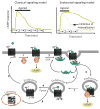
Figure 1
Figure 1. Classical versus endosomal signaling models of GPCR
Activation and desensitization of a cAMP response mediated by GPCR–GS systems proceed through a succession of biochemical and cellular events that initially take place at the cell membrane and result in the induction, propagation and termination of the second messenger molecule (steps 1–6). In the classical model, GPCR–G protein systems are only active on the cell surface and internalize to be degraded and/or replaced by newly synthesized GPCRs. In the new model, GS and cAMP signaling can continue after internalization of ligand–GPCR complexes in endosomes (step 3′). The figure is based on ref. . Arr, arrestin; Ppase, protein phosphatase.
Figure 2
Figure 2. Regulation of endosomal GPCR signaling
Proposed model of sustained cAMP signaling and its regulation. Left, PTH-activated PTHR generates cAMP by activation of adenylate cyclases internalizes to early endosomes in a process that involves binding of β-arrestins. Activated PTHR is then maintained in early endosomes by arrestin binding, where arrestin-mediated activation of ERK1/2 signaling causes inhibition of phosphodiesterases (PDEs) and permits sustained cAMP signaling. Right, binding of PTHR and retromer (blue) causes sorting of the receptor to retrograde trafficking domains. Generation of cAMP is stopped after PTHR–retromer binding in the retrograde domain and retromer-mediated PTHR traffic to the Golgi. Figure adapted from ref. .
Figure 3
Figure 3. Signaling models of GPCR
(a) Classical model. The ligand (L) binds the inactive state of a GPCR (R) and stabilizes its active form (R*), which then couples with heterotrimeric G proteins (Gαβγ) through a diffusion-controlled process (step 1). The L–R*–G complex, in turn, catalyzes GDP-GTP exchange on Gα, leading to dissociation the GTP-bound Gα(Gα-GTP) along with the Gβγ dimer from the receptor (step 2). In the case of GS, GαS-GTP activates ACs that catalyze the synthesis of cAMP from ATP (step 3). The hydrolysis of GTP to GDP causes the reassociation of GαS to Gβγ subunits and the termination of the cAMP production. In this model, the recruitment of β-arrestins mediate desensitization of G-protein signaling (step 4). (b) Noncanonical model, using PTHR as an example. (i) A long-lived PTH–PTHR–arrestin complex could contribute to sustained cAMP signaling by stabilizing an interaction with the active state of GS (i.e., the GTP-bound form of GS); (ii) alternatively, the interaction between the activated PTHR and Gβγ is stabilized by β-arrestins. After the first round of activation, the initial interaction between PTHR and Gs is bypassed such that, after hydrolysis of the GTP-bound form of GαS, free Gα-GDP directly reassociates with PTHR–Gβγ complexes to initiate a new cycle of G-protein activation. Arrestin stabilizes the G-protein cycle, resulting in prolonged cAMP production. Figure adapted from ref. .
Figure 4
Figure 4. Endosomal PTHR signaling: from bench to bedside
(a) Studies in cells led to the discovery that PTH, as opposed to PTHrP, sustains G-protein activity and cAMP production after PTHR internalization into early endosomes. (b) This observation is changing how we think about cellular signaling of the PTHR and is motivating the development of PTH analogs able to promote the endosomal cAMP signaling. One of them, LA-PTH, mediates a markedly prolonged cAMP signaling response in cells and prolonged hypercalcemic responses when injected into mice. (c) LA-PTH is now in preclinical development via the US National Institutes of Health BrIDGs program for eventual testing as a future treatment for hypoparathyroidism. Figure adapted from refs. , .
Figure 5
Figure 5. Differentiating PTHR conformations
(a) Competition radioligand binding isotherms. (b) Kinetics of radioligand dissociation. (c) Real-time kinetics of ligand dissociation measured by fluorescence resonance energy transfer (FRET). (d) Time course of cAMP in live cells measured by FRET. (e) Three-dimensional view of tetramethylrhodamine (TMR)-labeled peptides (red) and a PTHR N-terminally tagged with GFP (PTHRGFP, green) in live HEK-293 cells by confocal microscopy 30 min after ligand washout. Scale bars, 5 mm. The figure is based on ref. and is adapted from refs. , .

References

    1. Lefkowitz RJ. Seven transmembrane receptors: something old, something new. Acta Physiol (Oxf) 2007;190:9–19. - PubMed
    1. Vilardaga JP, Bunemann M, Krasel C, Castro M, Lohse MJ. Measurement of the millisecond activation switch of G protein–coupled receptors in living cells. Nat Biotechnol. 2003;21:807–812. - PubMed
    1. Nikolaev VO, Hoffmann C, Bunemann M, Lohse MJ, Vilardaga JP. Molecular basis of partial agonism at the neurotransmitter a2A-adrenergic receptor and Gi-protein heterotrimer. J Biol Chem. 2006;281:24506–24511. - PubMed
    1. Lohse MJ, et al. Optical techniques to analyze real-time activation and signaling of G-protein–coupled receptors. Trends Pharmacol Sci. 2008;29:159–165. - PubMed
    1. Vilardaga JP, Romero G, Feinstein TN, Wehbi VL. Kinetics and dynamics in the G protein–coupled receptor signaling cascade. Methods Enzymol. 2013;522:337–363. - PubMed

Publication types