

Mesenteric panniculitis: prevalence, clinicoradiological presentation and 5-year follow-up
- PMID: 25271412
- PMCID: PMC4243199
- DOI: 10.1259/bjr.20140451
Mesenteric panniculitis: prevalence, clinicoradiological presentation and 5-year follow-up
Abstract
Objective: To determine prevalence, clinicoradiological characteristics and outcome of patients with mesenteric panniculitis (MP) in a large hospital-based population.
Methods: Consecutive abdominal CT examinations of 3820 patients were evaluated for MP. Clinical characteristics, therapy and outcome of patients with MP were evaluated during a 5-year follow-up period. A matched pair analysis was performed to further investigate the relation between MP and malignancy.
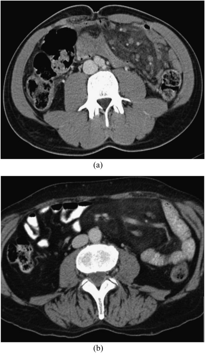
Results: 94 (2.5%) patients with MP were identified (mean age, 66.6 ± 11.2 years, 70.2% male). MP coexisted with malignancy (especially prostatic carcinoma) in 48.9% of patients, and this was slightly but significantly higher than in age- and sex-matched control patients (n = 188, 46.3%). In 48 patients, MP was presumed to be idiopathic. The most frequent presenting symptom was pain (54.3%). Laboratory findings revealed increased acute-phase reactants in half of the patients with MP. CT findings included increased density of mesenterial fat (mean, -56.8 ± 10.8 HU), fat ring sign, tumoural pseudocapsule and small soft-tissue nodules. Patients with MP (14.6%) developed significantly more malignancies during a 5-year follow-up than did the control group (6.9%). One patient was treated with prednisone without satisfactory response.
Conclusion: The prevalence of MP in this study was 2.5%. In most patients, radiologic features included increased mesenteric fat density, fat ring sign and small soft-tissue nodules. MP was associated with a significant higher prevalence of coexisting malignancies and a higher prevalence of future cancer development.
Advances in knowledge: A more accurate prevalence of MP on CT is demonstrated. An underlying malignancy may play a role.
Figures


References
-
- Emory TS, Monihan JM, Carr NJ, Sobin LH. Sclerosing mesenteritis, mesenteric panniculitis and mesenteric lipodystrophy: a single entity? Am J Surg Pathol 1997; 21: 392–8. - PubMed
-
- Kelly JK, Hwang WS. Idiopathic retractile (sclerosing) mesenteritis and its differential diagnosis. Am J Surg Pathol 1989; 13: 513–21. - PubMed
-
- Durst AL, Freund H, Rosenmann E, Birnbaum D. Mesenteric panniculitis: review of the leterature and presentation of cases. Surgery 1977; 81: 203–11. - PubMed
-
- Kipfer RE, Moertel CG, Dahlin DC. Mesenteric lipodystrophy. Ann Intern Med 1974; 80: 582–8. - PubMed
MeSH terms
LinkOut - more resources
Full Text Sources
Other Literature Sources
Medical
