Review
. 2015 Apr:84:188-97.
doi: 10.1016/j.addr.2014.09.008. Epub 2014 Sep 28.

Biomaterial-based delivery for skeletal muscle repair

Affiliations
Review

Biomaterial-based delivery for skeletal muscle repair

Christine A Cezar et al. Adv Drug Deliv Rev. 2015 Apr.

Abstract

Skeletal muscle possesses a remarkable capacity for regeneration in response to minor damage, but severe injury resulting in a volumetric muscle loss can lead to extensive and irreversible fibrosis, scarring, and loss of muscle function. In early clinical trials, the intramuscular injection of cultured myoblasts was proven to be a safe but ineffective cell therapy, likely due to rapid death, poor migration, and immune rejection of the injected cells. In recent years, appropriate therapeutic cell types and culturing techniques have improved progenitor cell engraftment upon transplantation. Importantly, the identification of several key biophysical and biochemical cues that synergistically regulate satellite cell fate has paved the way for the development of cell-instructive biomaterials that serve as delivery vehicles for cells to promote in vivo regeneration. Material carriers designed to spatially and temporally mimic the satellite cell niche may be of particular importance for the complete regeneration of severely damaged skeletal muscle.

Keywords: Cell therapy; Drug delivery; Microenvironmental cue; Muscle regeneration; Satellite cell; Synthetic niche; Tissue engineered muscle.


Figures

Figure 1
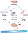
Figure 1. Impact of satellite cell isolation and manipulation on regenerative capacity
Following muscle biopsy and tissue dissociation, satellite cells are manipulated in different ways to obtain a desired cell population. The extent of manipulation generally determines the loss in regenerative potential that occurs before reinjection. Cells that are subjected to extensive in vitro culture (left) typically exhibit the greatest loss in regenerative potential and must be injected in high quantity. Alternatively, satellite cells that undergo a minimally intensive sorting procedure followed by immediate reinjection (middle) can be injected in lower quantities to obtain regenerative benefit. Satellite cells that are transplanted with their associated myofiber niche (right) demonstrate the greatest regenerative potential, and only a few fibers are required for significant muscle regeneration. Image used with permission [16].
Figure 2
Figure 2. Design criteria for a biomaterial-based synthetic satellite cell niche
Biophysical and biochemical cues can be used synergistically to create a synthetic satellite cell niche designed to optimize cell engraftment upon implantation. Biophysical cues, such as adhesive and mechanical signals, can dramatically affect cell survival and cell fate. For example, highly ordered material topography can be used to align myofibers and matrix stiffness can be used to promote self-renewal. Additionally, material carrier presentation of biochemical cues can lead to enhanced cell survival and participation in regeneration. Morphogens, cytokines, and angiogenic factors can be incorporated into a matrix through covalent coupling, physical entrapment, or ionic interactions in order to spatially and temporally control presentation. Key microenvironment cues are incorporated into an example synthetic satellite cell niche for cell transplantation.
Figure 3
Figure 3. Biomaterial-based approach to sustained cell delivery
A synthetic satellite cell niche can be designed to promote sustained cell delivery to injured muscle. Following implantation at a site of muscle damage, the engineered niche can provide protection from the harsh wound environment, and provide niche signals to promote cell activation leading to proliferation without terminal differentiation. Cell delivery to the injured area results from the subsequent outward migration of cells. Continued rounds of proliferation and outward migration lead to sustained cell delivery.
Figure 4
Figure 4. Tissue engineered skeletal muscle – bio-artificial muscle (BAM) formation
Tissue engineered skeletal muscle tissues are of potential clinical utility as therapeutic grafts to replace lost muscle tissue. In one approach, BAMs are created when an ECM/cell composite is allowed to contract against two anchor posts acting as artificial tendons. Although cells are initially seeded in the material with random orientation, they align and fuse to form myotubes with time. Following contraction of the material, a contractile, highly organized muscle tissue consisting of aligned myofibers is generated.

Similar articles

Cited by

References

    1. Juhas M, Bursac N. Engineering skeletal muscle repair. Curr Opin Biotechnol. 2013;24:880–886. - PMC - PubMed
    1. Turner NJ, Badylak SF. Regeneration of skeletal muscle. Cell Tissue Res. 2012;347:759–774. - PubMed
    1. Jarvinen TAHJ, Jarvinen TLN, Kaariainen M, Kalimo A, Jarvinen M. Muscle injuries - Biology and treatment. Am J Sports Med. 2005;33:745–764. - PubMed
    1. Rossi CA, Pozzobon M, De Coppi P. Advances in musculoskeletal tissue engineering Moving towards therapy. Organogenesis. 2010;6:167–172. - PMC - PubMed
    1. Tedesco FS, Cossu G. Stem cell therapies for muscle disorders. Curr Opin Neurol. 2012;25:597–603. - PubMed

MeSH terms

Substances