
Ibrutinib treatment ameliorates murine chronic graft-versus-host disease

Jason A Dubovsky et al. J Clin Invest. 2014 Nov.

Abstract

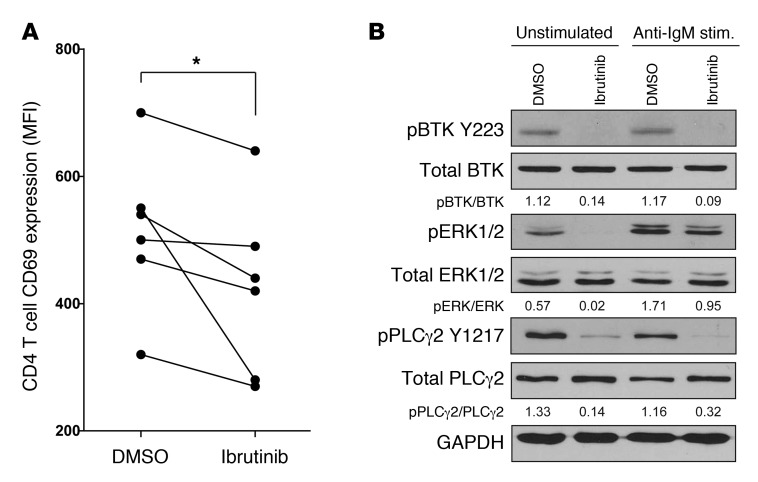
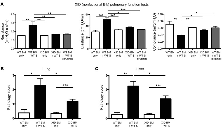
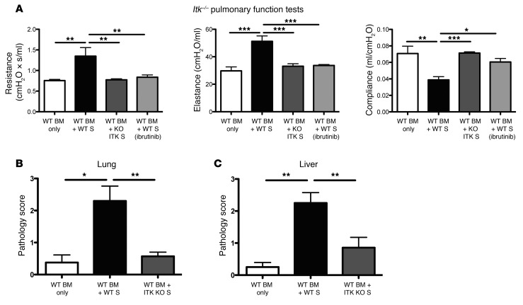
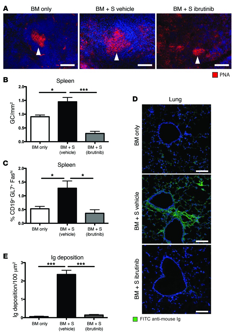
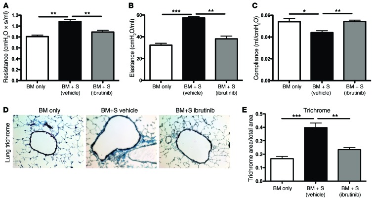
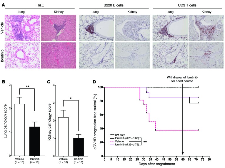
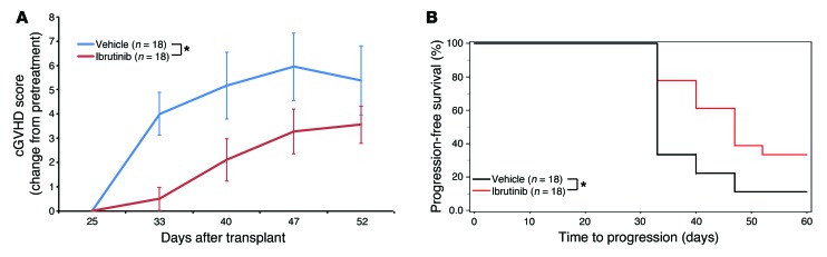
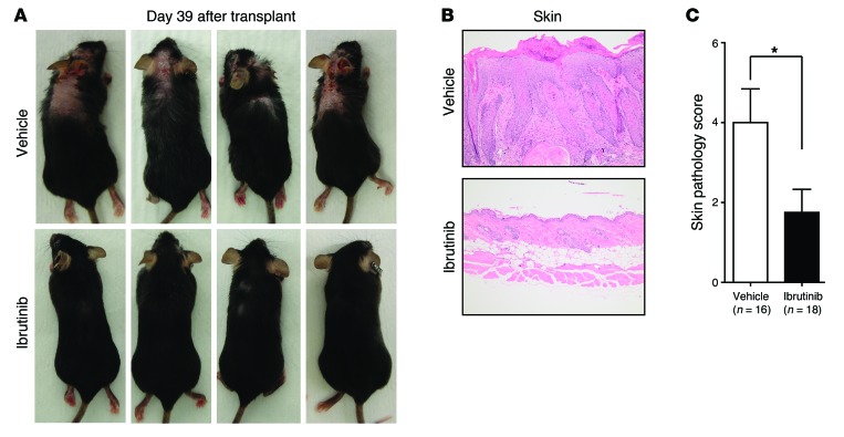
Chronic graft-versus-host disease (cGVHD) is a life-threatening impediment to allogeneic hematopoietic stem cell transplantation, and current therapies do not completely prevent and/or treat cGVHD. CD4+ T cells and B cells mediate cGVHD; therefore, targeting these populations may inhibit cGVHD pathogenesis. Ibrutinib is an FDA-approved irreversible inhibitor of Bruton's tyrosine kinase (BTK) and IL-2 inducible T cell kinase (ITK) that targets Th2 cells and B cells and produces durable remissions in B cell malignancies with minimal toxicity. Here, we evaluated whether ibrutinib could reverse established cGVHD in 2 complementary murine models, a model interrogating T cell-driven sclerodermatous cGVHD and an alloantibody-driven multiorgan system cGVHD model that induces bronchiolar obliterans (BO). In the T cell-mediated sclerodermatous cGVHD model, ibrutinib treatment delayed progression, improved survival, and ameliorated clinical and pathological manifestations. In the alloantibody-driven cGVHD model, ibrutinib treatment restored pulmonary function and reduced germinal center reactions and tissue immunoglobulin deposition. Animals lacking BTK and ITK did not develop cGVHD, indicating that these molecules are critical to cGVHD development. Furthermore, ibrutinib treatment reduced activation of T and B cells from patients with active cGVHD. Our data demonstrate that B cells and T cells drive cGVHD and suggest that ibrutinib has potential as a therapeutic agent, warranting consideration for cGVHD clinical trials.


Figures

Figure 8
Figure 8. Ibrutinib limits activation of T cells and B cells from patients with active cGVHD.
(A) Primary CD4+ T cells were isolated from patients with active cGVHD, pretreated with 1 μM ibrutinib (or DMSO), and stimulated using anti-CD3 for 6 hours. Graph shows the mean florescence intensity (MFI) for CD69 among CD4+ T cells for each patient. *P < 0.05. (B) B cells isolated from patients with cGVHD were pretreated with 1 μM ibrutinib and stimulated with anti-IgM for 45 minutes. Immunoblot analysis of BTK, ERK, and PLCγ2 was conducted. The densitometric quantification of activated proteins relative to total proteins is provided. Data are representative of 3 experiments on 3 separate patients.
Figure 7
Figure 7. Expression of BTK in donor-derived B cells is necessary for the development of BO.
(A) Day 60 PFTs from mice transplanted with low levels of WT T cells and either WT or XID (kinase inactive BTK) BM. (B) Pathologic scores in lung and (C) liver of day 60 transplanted mice. n = 5 mice/group from 2 independent experiments. *P < 0.05; **P < 0.01; ***P < 0.001.
Figure 6
Figure 6. Development of BO is dependent on ITK expression in donor mature T cells.
(A) Day 60 PFTs from mice transplanted with WT BM and low numbers of either WT T cells or ITK-deficient T cells. (B) Pathologic scores in lung and (C) liver of day 60 transplanted mice. n = 5 mice/group in 2 independent experiments. *P < 0.05; **P < 0.01; ***P < 0.001.
Figure 5
Figure 5. GC reactions and pulmonary immunoglobulin deposition are reduced with administration of ibrutinib.
(A) GCs were imaged by staining 6-μm spleen sections with PNA conjugated to rhodamine (red) and DAPI (blue). (B) GC area (GC/mm2) was calculated from PNA-stained immunofluorescent images for each animal. The average area for each cohort is displayed. Error bars indicate SEM. (C) Splenocytes were purified from transplanted mice on day 60, and frequency of GC B cells was quantified. (D) 6-μm lung sections from day 60 transplanted mice were stained with anti-mouse Ig conjugated to FITC (green) and DAPI (blue) and (E) quantified with Adobe Photoshop CS3. Representative data from 3 independent experiments. *P < 0.05; ***P < 0.001. All measurements were conducted on day 60 after HSCT. Scale bars: 100 μm.
Figure 4
Figure 4. Collagen deposition and pulmonary function are improved in a murine model of bronchiolitis obliterans.
(AC) PFTs were performed at day 60 after transplant on anesthetized animals. Animals (n = 4/group) were artificially ventilated and (A) resistance, (B) elastance, and (C) compliance were measured as parameters of distress in lung function in animals receiving 5 × 10⁶ splenocytes (S) in addition to BM. Error bars indicate SEM. (D and E) Collagen deposition within pulmonary tissues was determined with a Masson trichrome staining kit; blue indicates collagen deposition. (D) Representative images of collagen deposition observed in each treatment cohort (n = 8). Blue staining represents Masson trichrome–stained collagen. Original magnification, ×200. (E) Quantification of collaged deposition (n = 8) as a ratio of blue area to total area of tissue was performed with the analysis tool in Photoshop CS3. Representative data from 3 independent experiments. *P < 0.05; **P < 0.01; ***P < 0.001.
Figure 3
Figure 3. Ibrutinib therapy prevents autoimmune injury in a T cell–dependent model of cGVHD.
(A) Representative images from H&E-, B220-, or CD3-stained lung and kidney tissues from mice sacrificed at day 125 after HSCT from 6 mice/group. Images were taken by a trained veterinary pathologist who was blinded to animal cohorts. Original magnification, ×200. (B) Blinded pathologic analysis of H&E-stained lung tissues obtained from cGVHD cohorts (18 vehicle and 18 ibrutinib). Lymphohistiocytic infiltration was graded on a 0 to 4 scale for each animal. (C) Blinded pathologic analysis of H&E-stained kidney tissues obtained from cGVHD cohorts. Portal hepatitis and vasculitis were graded on a 0 to 4 scale for each animal. *P < 0.05; **P < 0.01. (D) Kaplan-Meier plot of cGVHD progression-free survival in an independent experiment aimed to determine sustained benefits from continued ibrutinib therapy. During the course of the experiment, ibrutinib was withdrawn on day 60 from animals in the Ibrutinib (day 25 to day 60) cohort. **P < 0.001.
Figure 2
Figure 2. Ibrutinib inhibits autoimmune manifestations of cGVHD.
(A) Weekly blinded analysis of cGVHD external metrics including weight, posture, vitality, mobility, coat, and skin in all mice from 2 independent experiments (18 vehicle and 18 ibrutinib) (Supplemental Table 1). All cGVHD scores were corrected for individual scores at the beginning of treatment (day 25). Error bars indicate SEM. *P < 0.01. (B) Kaplan-Meier plot of cGVHD progression–free survival. Data are derived from 2 independent experiments. Progression is defined as a greater than 2-point increase in day 25 cGVHD score (Supplemental Table 1) *P < 0.01.
Figure 1
Figure 1. Scleroderma and skin manifestations of cGVHD are alleviated by ibrutinib therapy.
At day 25 after HSCT, a total of 18 mice (from 2 independent experiments) were randomly assigned to ibrutinib (25 mg/kg/d), 18 to vehicle, and 11 to cyclosporine (10 mg/kg/d). Sclerodermatous lesions, hair loss, hunched posture, and gaunt appearance are characteristic visual indicators of cGVHD in this model. (A) Representative visual analysis of 4 randomly selected mice at day 39 after HSCT. (B) H&E-stained skin preparations of sclerodermatous skin lesions showing levels of dermal fibrosis, epidermal hyperplasia, serocellular crusting, erosion, and lymphohistiocytic infiltration, consistent with cGVHD. Original magnification, ×200. (C) Pathologic cGVHD involvement of the skin was independently assessed on a scale from 0 to 8 for each mouse. Cohort averages are displayed. *P < 0.05. Error bars indicate SEM.

References

    1. Baird K, Pavletic SZ. Chronic graft versus host disease. Curr Opin Hematol. 2006;13(6):426–435. doi: 10.1097/01.moh.0000245689.47333.ff. - DOI - PubMed
    1. Lee SJ, Vogelsang G, Flowers ME. Chronic graft-versus-host disease. Biol Blood Marrow Transplant. 2003;9(4):215–233. doi: 10.1053/bbmt.2003.50026. - DOI - PubMed
    1. Pidala J, et al. Patient-reported quality of life is associated with severity of chronic graft-versus-host disease as measured by NIH criteria: report on baseline data from the Chronic GVHD Consortium. Blood. 2011;117(17):4651–4657. doi: 10.1182/blood-2010-11-319509. - DOI - PMC - PubMed
    1. Arai S, et al. Global and organ-specific chronic graft-versus-host disease severity according to the 2005 NIH Consensus Criteria. Blood. 2011;118(15):4242–4249. doi: 10.1182/blood-2011-03-344390. - DOI - PMC - PubMed
    1. Holler E. Risk assessment in haematopoietic stem cell transplantation: GvHD prevention and treatment. Best Pract Res Clin Haematol. 2007;20(2):281–294. doi: 10.1016/j.beha.2006.10.001. - DOI - PubMed

Publication types

MeSH terms