. 2014 Oct 1;9(10):e108907.
doi: 10.1371/journal.pone.0108907. eCollection 2014.

Pre- and postovulatory aging of murine oocytes affect the transcript level and poly(A) tail length of maternal effect genes

Affiliations

Pre- and postovulatory aging of murine oocytes affect the transcript level and poly(A) tail length of maternal effect genes

Debora Dankert et al. PLoS One. .

Abstract

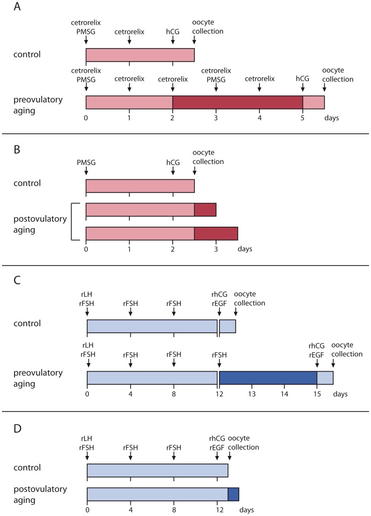
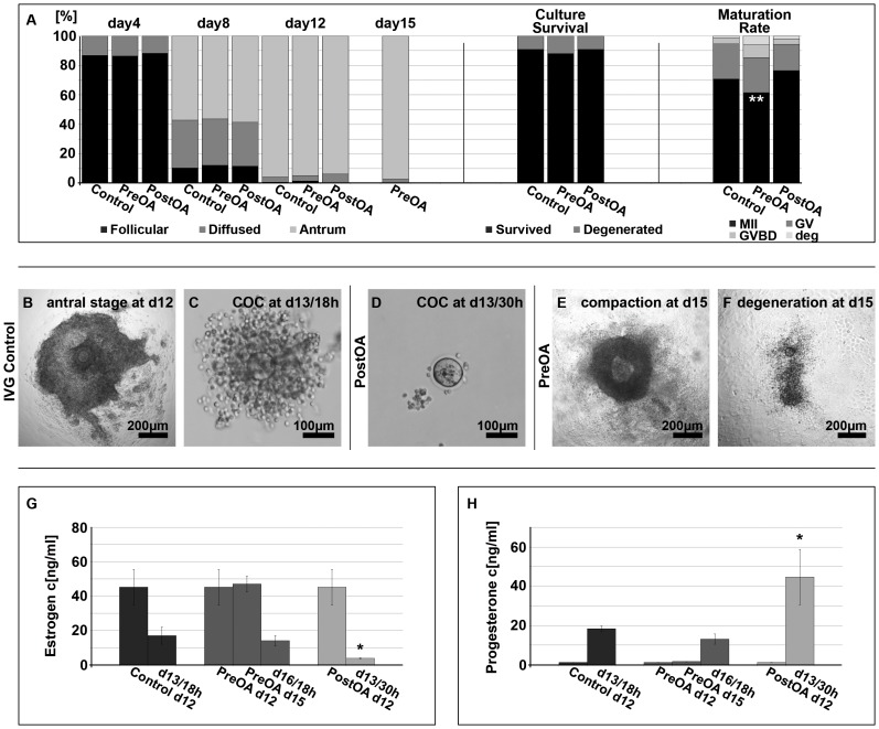
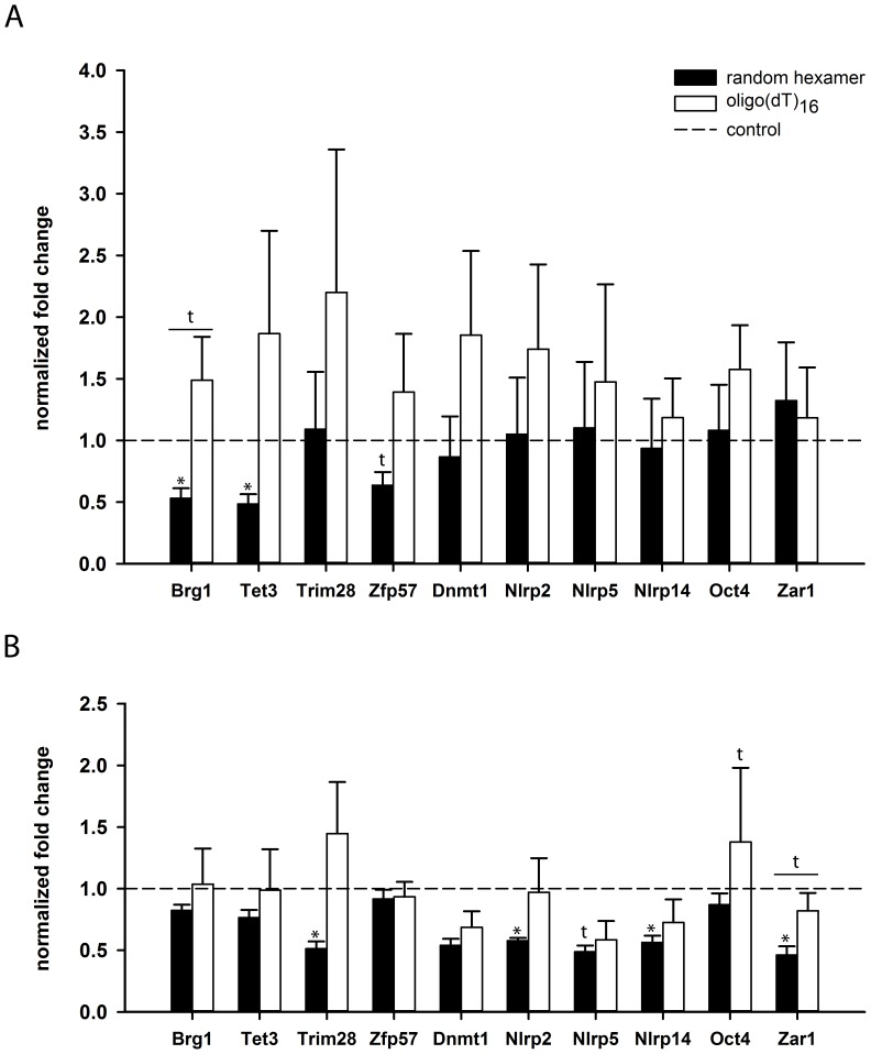
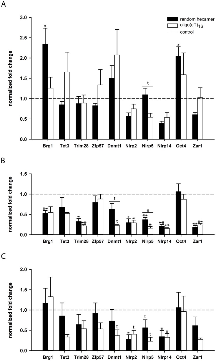
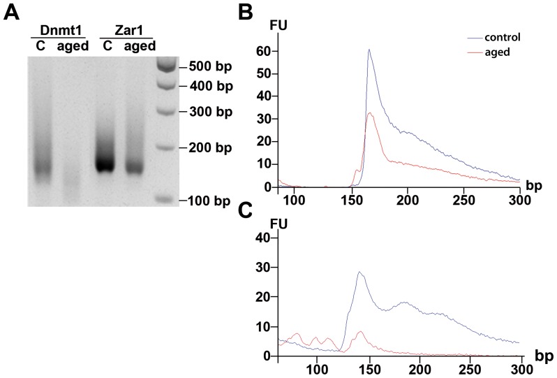
Maternal effect genes code for oocyte proteins that are important for early embryogenesis. Transcription in oocytes does not take place from the onset of meiotic progression until zygotic genome activation. During this period, protein levels are regulated posttranscriptionally, for example by poly(A) tail length. Posttranscriptional regulation may be impaired in preovulatory and postovulatory aged oocytes, caused by delayed ovulation or delayed fertilization, respectively, and may lead to developmental defects. We investigated transcript levels and poly(A) tail length of ten maternal effect genes in in vivo- and in vitro- (follicle culture) grown oocytes after pre- and postovulatory aging. Quantitative RT-PCR was performed using random hexamer-primed cDNA to determine total transcript levels and oligo(dT)16-primed cDNA to analyze poly(A) tail length. Transcript levels of in vivo preovulatory-aged oocytes remained stable except for decreases in Brg1 and Tet3. Most genes investigated showed a tendency towards increased poly(A) content. Polyadenylation of in vitro preovulatory-aged oocytes was also increased, along with transcript level declines of Trim28, Nlrp2, Nlrp14 and Zar1. In contrast to preovulatory aging, postovulatory aging of in vivo- and in vitro-grown oocytes led to a shortening of poly(A) tails. Postovulatory aging of in vivo-grown oocytes resulted in deadenylation of Nlrp5 after 12 h, and deadenylation of 4 further genes (Tet3, Trim28, Dnmt1, Oct4) after 24 h. Similarly, transcripts of in vitro-grown oocytes were deadenylated after 12 h of postovulatory aging (Tet3, Trim28, Zfp57, Dnmt1, Nlrp5, Zar1). This impact of aging on poly(A) tail length may affect the timed translation of maternal effect gene transcripts and thereby contribute to developmental defects.


Conflict of interest statement

Competing Interests: The authors have declared that no competing interests exist.

Figures

Figure 1
Figure 1. Timeline of pre- and postovulatory aging in vivo (A, B) and in vitro (C, D).
A, B) For in vivo maturation of oocytes, follicle maturation was stimulated by PMSG on day 0. Ovulation was induced by hCG 48 h later. Control oocytes were collected from the ampullae the next morning. For preovulatory in vivo aging, ovulation was delayed by the GnRH antagonist cetrorelix for 3 d (A). Oocytes for postovulatory aging were collected at the same time as controls and cultured in M2 medium for further 12 or 24 h (B). C, D) For in vitro growth and maturation of oocytes, preantral follicles were cultured for 12 d in the presence of rLH and rFSH to the antral follicle stage. Ovulation was induced with rhCG/rEGF and control oocytes were collected after 18 h. To obtain preovulatory-aged oocytes, ovulation was induced with rhCG/rEGF on day 15 of follicle culture instead of day 12 (C). For postovulatory aging, ovulation was triggered with rhCG/rEGF and oocytes were incubated for additional 12 h before collection (D).
Figure 2
Figure 2. Follicle morphology, morphokinetics, and hormone concentrations in conditioned medium of preantral follicle culture.
A) Follicle characteristics, culture survival and maturation of control (n = 1230), preovulatory-aged (PreOA; n = 411) and postovulatory-aged (PostOA; n = 613) oocytes. B-D) Antral stage follicle grown in vitro for 12 d (B) and cumulus-oocyte complexes on day 13 after in vitro ovulation in control oocytes (C) and after postovulatory aging (D) for 12 h. E, F) Altered granulosa cell characteristics after preovulatory aging at day 15 of culture; follicles with an increased accumulation of mural granulosa cells and an apparent follicle compaction (E), and a degenerating follicle with dispersed granulosa cells and a nearly denuded oocyte from day 15 of culture (F). G) Estrogen and (H) progesterone levels (mean ± SEM) in conditioned culture medium prior to and past hormonal stimulation by rhCG/rEGF in the different experimental groups (* P<0.05, ** P<0.01).
Figure 3
Figure 3. Expression levels and poly(A) content of ME genes in preovulatory-aged oocytes.
The normalized fold change (mean ± SEM) of preovulatory-aged oocytes compared to control oocytes (dotted line) of total transcript (black bars) and polyadenylated transcript (white bars) is shown. A) After preovulatory in vivo aging, oocytes show a significant decline in total transcript levels for Brg1 and Tet3. Comparison of total with polyadenylated transcript levels reveals that poly(A) content of ME gene mRNA tends to increase during preovulatory in vivo aging. B) Total transcript amounts of Trim28, Nlrp2, Nlrp14 and Zar1 decreased significantly after preovulatory aging in vitro. A similar trend was observed for Nlrp5. Several genes investigated showed a tendency towards a relative increase in poly(A) content compared to total transcript levels, which was most evident for Zar1 (t: P<0.10, * P<0.05).
Figure 4
Figure 4. Expression levels and poly(A) content of ME genes in postovulatory-aged oocytes.
The normalized fold change (mean ± SEM) of postovulatory-aged oocytes compared to control oocytes (dotted line) of total transcript (black bars) and polyadenylated transcript (white bars) is shown. In vivo-grown oocytes were aged for 12 h (A) and 24 h (B). After 12 h of postovulatory aging, the stronger reduction of polyadenylated transcript in comparison to total transcript levels indicates a reduced poly(A) tail length for Nlrp5. After 24 h, postovulatory aging results in a considerable decline of overall transcript amount and a decline in poly(A) content for most of the genes investigated. This decline in poly(A) content was significant for Nlpr5 and showed a trend for Dnmt1. C) Poly(A) content of in vitro-grown oocytes tended to decline for eight of the ten genes investigated already after 12 h of postovulatory aging, (t: P<0.10, * P<0.05, ** P<0.01).
Figure 5
Figure 5. Quantification of poly(A) tail length for Dnmt1 and Zar1 by ePAT.
24 h postovulatory-aged, in vivo-grown oocytes were analyzed by extension poly(A) test (ePAT). A) Gel electrophoresis of the product shows a decrease of poly(A) tail length for Dnmt1 in aged oocytes compared to controls, whereas poly(A) tail length of Zar1 remains widely stable. These results were quantified by capillary electrophoresis for Zar1 (B) and Dnmt1 (C). Indicated is the fluorescence intensity (FU) of amplicon lengths (in base pairs) for aged oocytes (red line) and controls (blue line).

References

    1. Kang MK, Han SJ (2011) Post-transcriptional and post-translational regulation during mouse oocyte maturation. BMB Rep 44: 147–157. - PubMed
    1. Clarke HJ (2012) Post-transcriptional control of gene expression during mouse oogenesis. Berlin Heidelberg: Springer-Verlag. - PubMed
    1. Weill L, Belloc E, Bava FA, Mendez R (2012) Translational control by changes in poly(A) tail length: recycling mRNAs. Nat Struct Mol Biol 19: 577–585. - PubMed
    1. Beilharz TH, Preiss T (2007) Widespread use of poly(A) tail length control to accentuate expression of the yeast transcriptome. RNA 13: 982–997. - PMC - PubMed
    1. McGrew LL, Dworkin-Rastl E, Dworkin MB, Richter JD (1989) Poly(A) elongation during Xenopus oocyte maturation is required for translational recruitment and is mediated by a short sequence element. Genes Dev 3: 803–815. - PubMed

Publication types

LinkOut - more resources