. 2014 Oct 1;9(10):e108946.
doi: 10.1371/journal.pone.0108946. eCollection 2014.

Structural insights into RNA polymerase recognition and essential function of Myxococcus xanthus CdnL

Affiliations

Structural insights into RNA polymerase recognition and essential function of Myxococcus xanthus CdnL

Aránzazu Gallego-García et al. PLoS One. .

Abstract

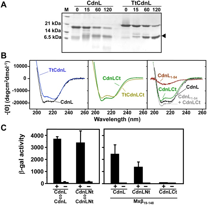
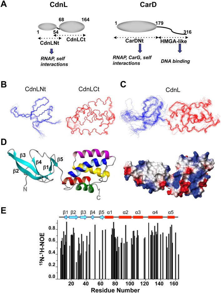
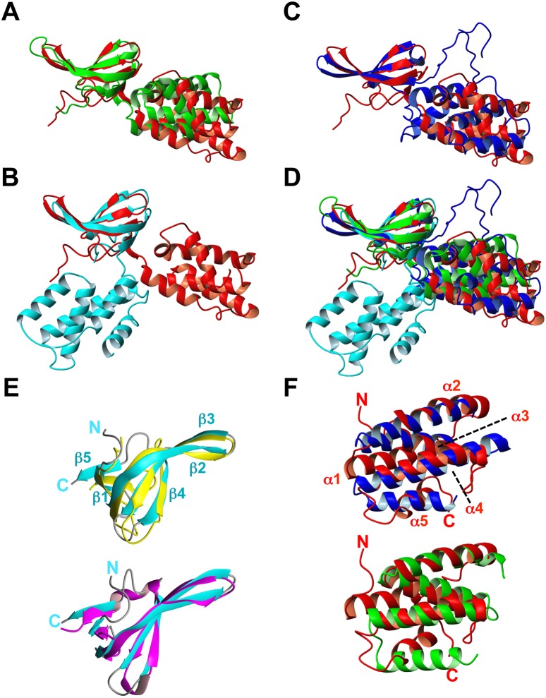
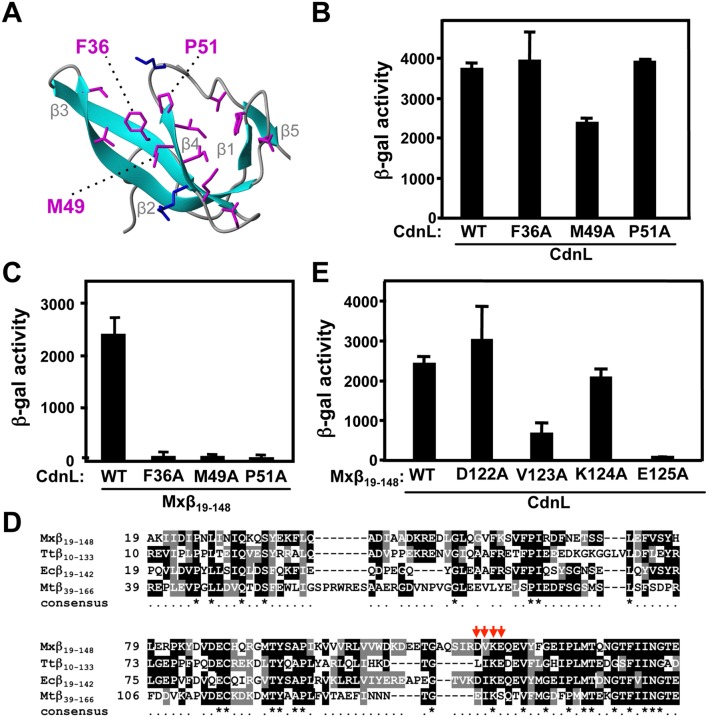
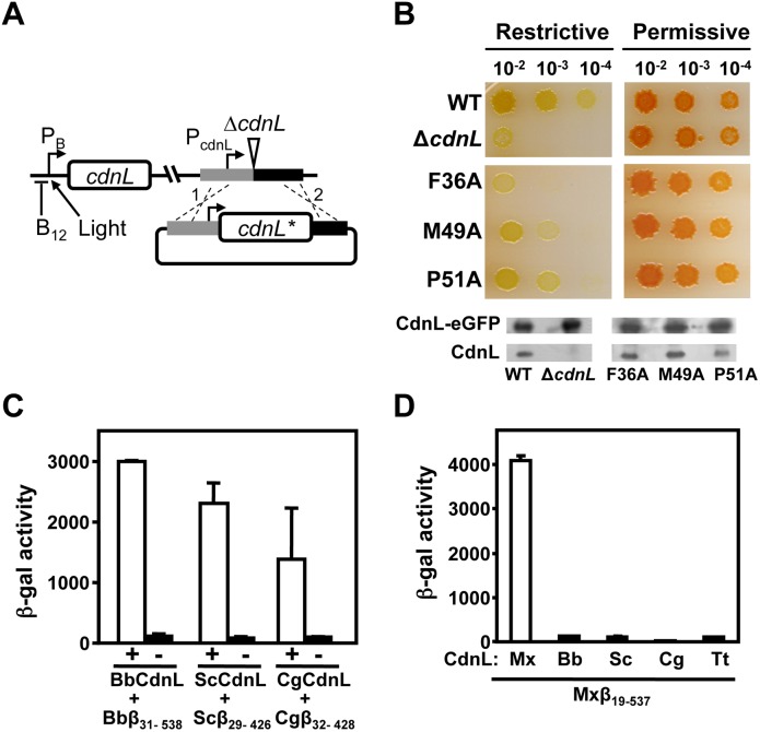
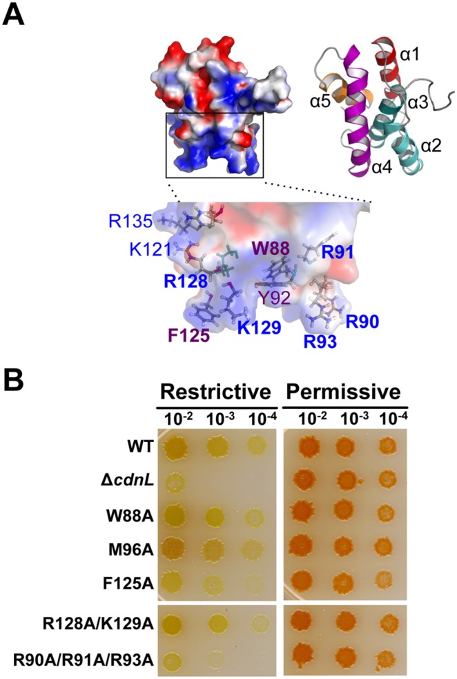
CdnL and CarD are two functionally distinct members of the CarD_CdnL_TRCF family of bacterial RNA polymerase (RNAP)-interacting proteins, which co-exist in Myxococcus xanthus. While CarD, found exclusively in myxobacteria, has been implicated in the activity of various extracytoplasmic function (ECF) σ-factors, the function and mode of action of the essential CdnL, whose homologs are widespread among bacteria, remain to be elucidated in M. xanthus. Here, we report the NMR solution structure of CdnL and present a structure-based mutational analysis of its function. An N-terminal five-stranded β-sheet Tudor-like module in the two-domain CdnL mediates binding to RNAP-β, and mutations that disrupt this interaction impair cell growth. The compact CdnL C-terminal domain consists of five α-helices folded as in some tetratricopeptide repeat-like protein-protein interaction domains, and contains a patch of solvent-exposed nonpolar and basic residues, among which a set of basic residues is shown to be crucial for CdnL function. We show that CdnL, but not its loss-of-function mutants, stabilizes formation of transcriptionally competent, open complexes by the primary σA-RNAP holoenzyme at an rRNA promoter in vitro. Consistent with this, CdnL is present at rRNA promoters in vivo. Implication of CdnL in RNAP-σA activity and of CarD in ECF-σ function in M. xanthus exemplifies how two related members within a widespread bacterial protein family have evolved to enable distinct σ-dependent promoter activity.


Conflict of interest statement

Competing Interests: The authors have declared that no competing interests exist.

Figures

Figure 1
Figure 1. CdnL domain analysis.
(A) Limited proteolysis of CdnL and TtCdnL. Aliquots of the reaction mix (1∶100 w/w subtilisin:protein) at 30°C were withdrawn at the times (in min) indicated on top and analyzed in Coomassie-stained 15% SDS-PAGE gels (lane “M”: molecular weight markers). Arrowhead on the right points to the subtilisin-resistant fragment, which mass spectrometry and N-terminal sequencing identified as the ∼100-residue C-terminal region in both proteins. Note the slower mobility of TtCdnL, whose size (164 residues) is the same as CdnL. (B) Far UV-CD spectra of CdnL, TtCdnL, and their indicated fragments. (C) BACTH analysis of the interactions of CdnL and its domains showing reporter lacZ expression in E. coli transformed with plasmids pKT25 and pUT18 bearing cdnL or the gene for CdnLNt (left panel); or with a pUT18C construct of the gene for Mxβ19–148 and pKT25 with cdnL or its indicated fragments (right panel). pKT25 without insert was used in negative controls (“−”).
Figure 2
Figure 2. NMR solution structures of CdnL and its domains.
(A) Schematic comparing CdnL and CarD domain architectures. Structured domains are shown as ellipsoids, intrinsically unstructured domains as wavy lines, and known interactions of each domain are indicated. Residues delimiting the CdnL and CarD domains are numbered. Note that CdnL resembles CarDNt in size and sequence, and lacks the HMGA-like DNA binding domain that is present in CarD. NMR structures of CdnLNt, CdnLCt, and full-length CdnL were determined in this study. (B) Superposition of the backbone traces for the 20 final NMR structures for CdnLNt (blue), CdnLCt (red), as indicated. (C) Same as in (B) for full length CdnL. (D) Average NMR solution structure of native CdnL in ribbon (left) and electrostatic surface representations (right). In the ribbon structure, the five β-strands (in cyan) of the CdnLNt domain are indicated, and the CdnLCt α-helices are colored red (α1), magenta (α2), yellow (α3), blue (α4) and green (α5). (E) Steady state backbone 15N-1H NOE (from the ratio of cross-peak intensities with and without 1H saturation at pH 7.0 and 25°C) plotted versus residue number obtained for 1 mM 15N, 13C-labeled CdnL. Errors for the heteronuclear NOE data were estimated to be ≤5% based on average noise levels in the NMR spectra.
Figure 3
Figure 3. Structural comparisons with M. xanthus CdnL NMR structure.
Backbone overlay of the CdnL NMR structure (red) onto the crystal structures of (A): TtCdnL (green; PDB: 4L5G); (B) one unit in the MtCdnL domain-swapped dimer (blue; PDB: 4ILU); (C) MtCdnL in complex with the RNAP β-lobe domain (cyan; PDB: 4KBM); (D) all four structures. The overlay shown was generated with optimal (maximum) superposition of the N-terminal domains of each structure. (E) Backbone overlay of CdnLNt (cyan; left) onto: (top) TtCdnLNt (gold; PDB ID: 2lqK); (bottom) E. coli TRCF-RID (magenta; PDB ID: 2eyq). (F) Backbone overlay of CdnLCt (red) onto the TPR domains of: (top) FK506-binding protein (blue; PDB ID: 1kt0); (bottom) CadCpd (green; PDB ID: 3ly8). Structures were generated with MOLMOL.
Figure 4
Figure 4. Mutational analysis of CdnL interaction with RNAP-β.
(A) Native CdnLNt structure showing residue side chains that may contact RNAP-β, with the three labeled tested by mutational analysis (see text). (B) BACTH analysis showing that the F36A, M49A and P51A CdnL variants self-interact in cells with the pKT25 and pUT18 constructs of wild type or mutant cdnL, as indicated. (C) BACTH analysis of the interaction with Mxβ19–148 (in pUT18C) of CdnL F36A, M49A, or P51A mutants in pKT25. (D) Sequence alignment of the M. xanthus RNAP-β segment, Mxβ19–148 (NCBI code, YP_631280.1) and its equivalents in T. thermophilus, Ttβ10–133 (WP_014630291), in E. coli, Ecβ19–142 (P0A8V2), and in M. tuberculosis (CAB09390). Residues shaded black if identical, or grey if similar, in at least two of the aligned sequences. An asterisk in the consensus line below indicates conservation in all four sequences. Red arrows point to RNAP-β residues analyzed by site-directed mutagenesis in this study. (E) BACTH analysis of the interaction of Mxβ19–148 (WT) or its indicated mutants in pUT18C versus wild-type CdnL in pKT25.
Figure 5
Figure 5. Consequences of disrupting CdnL interaction with RNAP in vivo.
(A) Scheme of the strategy employed to check for cdnL complementation in M. xanthus. A pMR2873 construct with the required cdnL allele (cdnL*) under PcdnL control and DNA segments flanking cdnL upstream (grey) and downstream (black) in the genome, was introduced into strain MR1467, which bears the ΔcdnL allele and a copy of cdnL at a heterologous site under the control of the photoinducible PB promoter (expressed in the light but repressed in the presence of B12 in the dark). Merodiploids resulting from plasmid integration by recombination at either “1” or “2” would constitutively express cdnL* from PcdnL and conditionally express the wild-type allele from PB at a heterologous site. (B) Complementation analyses in M. xanthus with the F36A, M49A and P51A CdnL mutants. CTT/B12 plates that were spotted with 5 µl of liquid cultures (OD550 ∼1) grown under permissive conditions and diluted as indicated, and then incubated for 2 days at 33°C under restrictive (dark and B12) or permissive (light, hence the red color) conditions. “WT” is the positive control derived from using pMR2873 with wild-type cdnL, and “ΔcdnL” is the recipient strain MR1467. Western blots (using polyclonal anti-CdnL antibodies) of M. xanthus cell extracts from strains expressing each CdnL variant and in which CdnL-eGFP supplied the essential CdnL function, as described in the text. (C) BACTH analysis of the interaction of CdnL homologs (BbCdnL, ScCdnL, and CgCdnL) with Bbβ31–538, Scβ29–426, and Cgβ32–428 fragments, respectively. “+” below the open bars indicates that both members of the pair are present, while “−” is the negative control (without the indicated CdnL homolog). We have shown the interaction of TtCdnL with its cognate β-fragment elsewhere . (D) BACTH analysis of the interaction of BbCdnL, ScCdnL, CgCdnL or TtCdnL with M. xanthus Mxβ19–537.
Figure 6
Figure 6. Consequences in vivo of mutating the CdnLCt solvent-exposed basic-hydrophobic patch.
(A) CdnLCt electrostatic surface (left) and ribbon representations (right; each helix is labeled and colored differently) generated using PyMOL. The zoom shows sidechains (as sticks) of the labeled nonpolar and basic residues composing the solvent-exposed basic-hydrophobic patch. (B) Complementation analyses in M. xanthus of the CdnL W88A, M96A, F125A, R128A/K129A, and R90A/R91A/R93A mutants versus wild-type CdnL (WT) and ΔcdnL, as in Figure 5B.
Figure 7
Figure 7. CdnL favors RPo formation and transcription from σA-dependent promoters.
(A) Promoter sequences for the σA-dependent P4rrnD and PB, indicating the -35 and -10 elements (in boxes) and the transcription start site. For comparison, the ECF-σ CarQ-dependent PQRS promoter is also included, with the conserved motifs at the -35 and -10 regions underlined. (B) ChIP-qPCR analysis of CdnL enrichment at P4rrnD, PB and PQRS in M. xanthus carried out as described in the text. The mean and standard of three independent experiments are shown. (C)–(E) EMSA of the binding of the 32P-labeled 130-bp PB or 151-bp P4rrnD DNA probes to 130 nM RNAP-σA holoenzyme, alone or in the presence of 10 µM of CdnL, CdnLNt, CdnLCt, or CarDNt (C), or a given CdnL variant (D, E), as indicated. After incubation for 30-min at 37°C, the complexes formed were challenged by adding 1 µg heparin. Note that any alteration in the migration of the shifted complexes due to CdnL binding to RNAP was not discernible, possibly because CdnL is considerably smaller than RNAP. (F) Single round, run-off in vitro transcription from P4rrnD (see Materials and methods) with 130 nM RNAP-σA alone or with 10 µM CdnL (WT or mutant) followed by heparin challenge (1 µg) and then addition of the labeled NTP mix to initiate transcription. In CF, representative data from three or more experiments are shown.

Similar articles

Cited by

References

    1. Lee DJ, Minchin SD, Busby SJ (2012) Activating transcription in bacteria. Annu Rev Microbiol 66: 125–152. - PubMed
    1. Haugen SP, Ross W, Gourse RL (2008) Advances in bacterial promoter recognition and its control by factors that do not bind DNA. Nat Rev Microbiol 6: 507–519. - PMC - PubMed
    1. Lemke JJ, Sanchez-Vazquez P, Burgos HL, Hedberg G, Ross W, et al. (2011) Direct regulation of Escherichia coli ribosomal protein promoters by the transcription factors ppGpp and DksA. Proc Natl Acad Sci U S A 108: 5712–5717. - PMC - PubMed
    1. García-Moreno D, Abellón-Ruiz J, García-Heras F, Murillo FJ, Padmanabhan S, et al. (2010) CdnL, a member of the large CarD-like family of bacterial proteins, is vital for Myxococcus xanthus and differs functionally from the global transcriptional regulator CarD. Nucleic Acids Res 38: 4586–4598. - PMC - PubMed
    1. Stallings CL, Stephanou NC, Chu L, Hochschild A, Nickels BE, et al. (2009) CarD is an essential regulator of rRNA transcription required for Mycobacterium tuberculosis persistence. Cell 138: 146–159. - PMC - PubMed

Publication types

MeSH terms

Substances

LinkOut - more resources