

A philosophy for CNS radiotracer design
- PMID: 25272291
- PMCID: PMC4204929
- DOI: 10.1021/ar500233s
A philosophy for CNS radiotracer design
Abstract
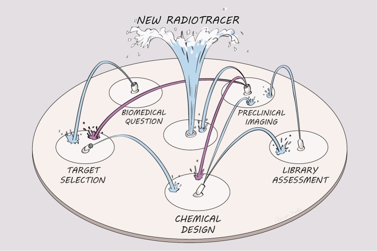
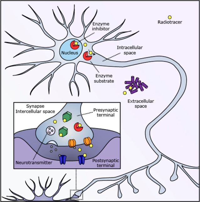
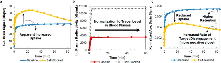
Decades after its discovery, positron emission tomography (PET) remains the premier tool for imaging neurochemistry in living humans. Technological improvements in radiolabeling methods, camera design, and image analysis have kept PET in the forefront. In addition, the use of PET imaging has expanded because researchers have developed new radiotracers that visualize receptors, transporters, enzymes, and other molecular targets within the human brain. However, of the thousands of proteins in the central nervous system (CNS), researchers have successfully imaged fewer than 40 human proteins. To address the critical need for new radiotracers, this Account expounds on the decisions, strategies, and pitfalls of CNS radiotracer development based on our current experience in this area. We discuss the five key components of radiotracer development for human imaging: choosing a biomedical question, selection of a biological target, design of the radiotracer chemical structure, evaluation of candidate radiotracers, and analysis of preclinical imaging. It is particularly important to analyze the market of scientists or companies who might use a new radiotracer and carefully select a relevant biomedical question(s) for that audience. In the selection of a specific biological target, we emphasize how target localization and identity can constrain this process and discuss the optimal target density and affinity ratios needed for binding-based radiotracers. In addition, we discuss various PET test-retest variability requirements for monitoring changes in density, occupancy, or functionality for new radiotracers. In the synthesis of new radiotracer structures, high-throughput, modular syntheses have proved valuable, and these processes provide compounds with sites for late-stage radioisotope installation. As a result, researchers can manage the time constraints associated with the limited half-lives of isotopes. In order to evaluate brain uptake, a number of methods are available to predict bioavailability, blood-brain barrier (BBB) permeability, and the associated issues of nonspecific binding and metabolic stability. To evaluate the synthesized chemical library, researchers need to consider high-throughput affinity assays, the analysis of specific binding, and the importance of fast binding kinetics. Finally, we describe how we initially assess preclinical radiotracer imaging, using brain uptake, specific binding, and preliminary kinetic analysis to identify promising radiotracers that may be useful for human brain imaging. Although we discuss these five design components separately and linearly in this Account, in practice we develop new PET-based radiotracers using these design components nonlinearly and iteratively to develop new compounds in the most efficient way possible.
Figures

References
-
- Lockwood A. H. Invasiveness in Studies of Brain Function by Positron Emission Tomography (PET). J. Cereb. Blood Flow Metab. 1985, 5, 487–489. - PubMed
-
- Eriksson L.; Kanno I. Blood Sampling Devices and Measurements. Med. Prog. Technol. 1991, 17, 249–257. - PubMed
-
- Zimmer L.; Luxen A. PET Radiotracers for Molecular Imaging in the Brain: Past, Present and Future. NeuroImage 2012, 61, 363–370. - PubMed
-
- Hargreaves R. J.; Rabiner E. A. Translational PET Imaging Research. Neurobiol. Dis. 2014, 61, 32–38. - PubMed
-
- CNS Radiotracer Table http://www.nimh.nih.gov/research-priorities/therapeutics/cns-radiotracer... (accessed Jun 17, 2014).
Publication types
MeSH terms
Substances
Grants and funding
LinkOut - more resources
Full Text Sources
Other Literature Sources
Research Materials
