. 2014 Nov;33(6):581-91.
doi: 10.1097/PGP.0000000000000095.

Characterization of the immune cell repertoire in the normal fallopian tube

Affiliations

Characterization of the immune cell repertoire in the normal fallopian tube

Laura Ardighieri et al. Int J Gynecol Pathol. 2014 Nov.

Abstract

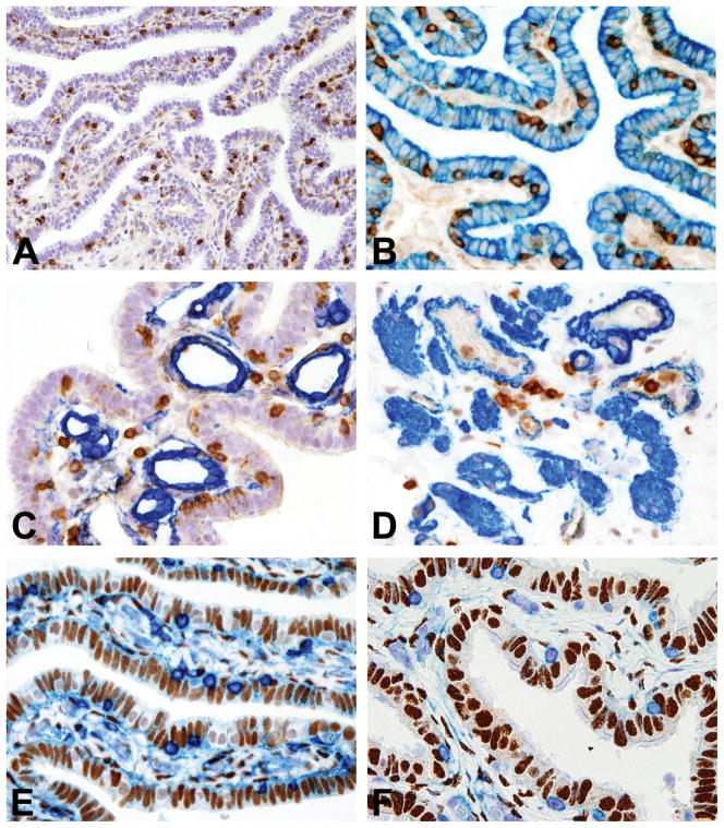
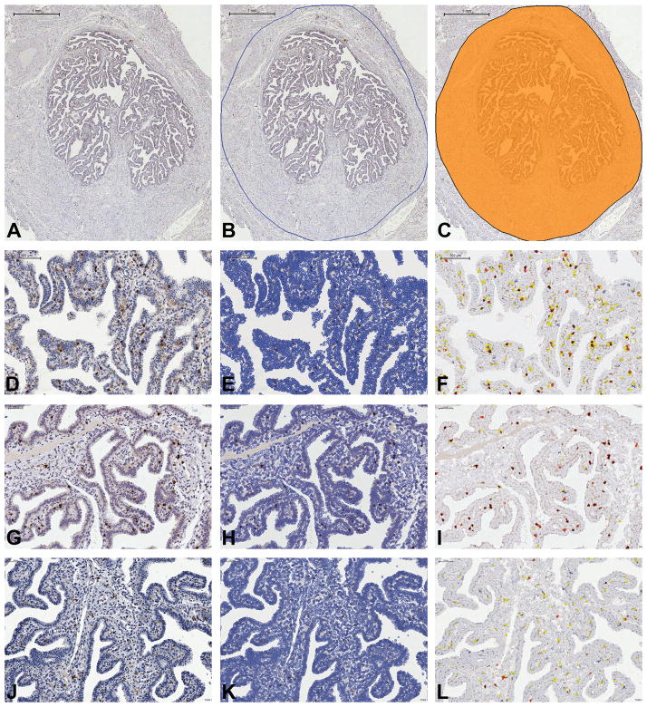
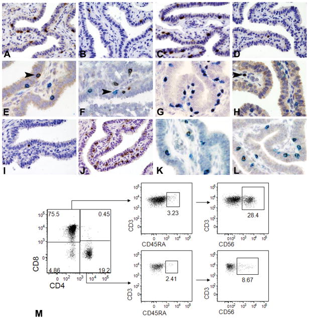
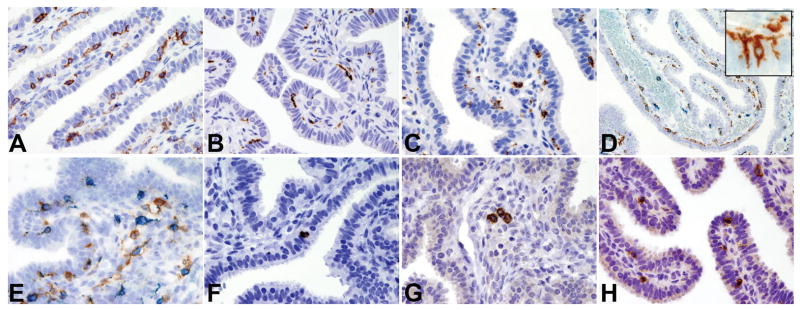
Recent studies implicating the fallopian tube as the site of putative precursors of ovarian serous carcinoma, and the hypothesis that injury, inflammation, and repair of the ovarian surface epithelium at the time of ovulation, may be contributing factors to ovarian carcinogenesis, prompted us to undertake a comprehensive analysis of the immune cells in the normal fallopian tube. Accordingly, the aim of this study was to provide a baseline for future studies exploring the relationship of inflammation with the early events of ovarian carcinogenesis by characterizing the immune cell repertoire in 13 normal human fallopian tubes, combining digital microscopy of immunostained slides and flow cytometry of fresh single-cell suspensions, with a panel of markers that identify the most important adaptive and innate immune cells. We found that CD45(+) leukocytes are regularly observed in the fallopian tube and are mainly composed of CD163(+) macrophages, CD11c dendritic cells, and CD8(+) T cells. In addition, there are minor populations of CD56(+) NK cells, CD4(+) T cells, CD20(+) B cells, TCRγδ(+) T cells, and, among dendritic cells, CD207(Langerin)(+) Langerhans cells. The cellular mapping that we performed indicates that the local immune system in the human fallopian tube is composed of a mixture of innate and adaptive immune cells, many of which are recognized as playing a role in cancer immune surveillance. This local immune system could provide a first line of defense against early precancerous lesions and could potentially be exploited for immune-based therapies.


Conflict of interest statement

Disclosure/Conflict of Interest

The authors declare absence of a conflict of interest.

Figures

Figure 1
Figure 1. Localization and hormone receptor expression of resident leucocyte populations in normal fallopian tube
Sections are from FFPE normal fallopian tube (FT) (A–F). A illustrates CD45+ immune cells in a FT isthmic section; B illustrates CD45(brown)/CK7(blue) double immunostaining showing CD45+ immune cells intermingled with CK7+ epithelial cells; C illustrates CD45(brown)/CD34(blue) double immunostaining showing CD45+ immune cells in close proximity to CD34+ vessels of the FT lamina propria; D illustrates CD45(brown)/smooth muscle actin (blue) double immunostaining showing CD45+ immune cells in close proximity to smooth muscle actin+ smooth muscle fibers/vessels of the FT muscular wall; E–F illustrate double immunostaining with CD45 (E–F, blue) and ER (brown, E) and PgR (brown, F) showing no expression of hormones receptors by immune cells. Sections are counterstained with Meyer’s haematoxylin. Magnification: 20× (A) and 40× (B–F).
Figure 2
Figure 2. Digital Microscopy analysis of the main immune cell populations in the normal fallopian tube (FT)
Sections are from FFPE normal fallopian tube and are digitalized by Aperio Scanscope and processed by Tissue Studio Definiens. A–C illustrate ROI (region of interest) selection: A illustrates the digital image of whole isthmic section of FT; B illustrates the manual selection of the ROI; C indicates the ROI (orange color) submitted to Tissue Studio Definiens for image analysis (Bar scale: A–C 1mm). D–F, G–I, J–L are matched native (D, G and J) and processed for nuclear identification (E, H and K) and signal detection (F, I and L; white cells are negative cells whereas yellow, orange and red cells correspond to positive cells with different signal intensity) images (Bar scale: D–F 100μm; G–L: 50 μm).
Figure 3
Figure 3. Cellular populations of the adaptive immune system in the normal fallopian tube
Sections from FFPE normal fallopian tube are analyzed by immunohistochemistry (A–L) whereas single cell suspension of fresh normal fallopian tube is analyzed by flow cytometry (M). A–D illustrate the main lymphoid populations detected in the intraepithelial compartment (IE) including CD3+ T-cells (A), CD20+ B-cells (B), CD8+ cytotoxic T-cells (C) and CD4+ T-helper cells (D). E illustrates CD3(blue)/CD56(brown) double immunostaining showing co-expression of CD56 NK marker by a CD3+ T-cells (arrow head). F illustrates CD3(brown)/CD56(blue) double immunostaining showing the presence of CD3+CD8+ T-cells (prevalent subtype) and CD3CD8+ cells (arrow head), likely representative of CD8+ NK subset. G illustrates CD3(blue)/T-bet (brown) double immunostaining showing the prevalence of Tbet+CD3+ T-cells. H illustrates CD3(blue)/Foxp3(brown, nuclear) double immunostaining showing absence of Foxp3+ regulatory T-cell in the IE compartment; arrow head indicates a single CD3+Foxp3+ regulatory T-cells in the lamina propria. I–J show comparison between IE TCRγδ+ (I) and TCRαβ+ T-cells (J). K–L illustrate double immunostaining with CD3 (K–L, blue) and Granzyme B (K, brown) and Perforin (L, brown) showing the presence of those cytotoxic molecules in CD3+ T-cells. Sections are counterstained with Meyer’s haematoxylin. Magnification: 40× (A–D, I–J) and 60× (E–H, K–L). M shows flow cytometry characterization of T lymphocytes on digested tubal tissue. Left plot: CD4/CD8 ratio; central and right plots: percentage of CD45RA+ and CD56+ cells, respectively, in the CD8+CD4 and CD4+CD8 gates (The panel is representative of three different samples).
Figure 4
Figure 4. Cellular populations of the innate immune system in the normal fallopian tube
Sections are from FFPE normal fallopian tube (A–H) and stained for CD11c (A), CD68 (B) and CD163 (C). D–E illustrate CD163(blue)/CD11c(brown) double immunostaining showing the presence respectively of CD11c+CD163 cells in the intraepithelial compartment (D; insert showing intraepithelial cytoplasmic projections of dendritic cells) and of CD11c+CD163+ cells admixed to CD11cCD163+ cells in the lamina propria (E); F illustrates CD207/Langerin+ intraepithelial Langerhans cell; G illustrates occasional CD303/BDCA2+ plasmacytoid dendritic cells in vessels of the lamina propria; H illustrates intraepithelial CD56+ NK cells. Sections are counterstained with Meyer’s haematoxylin. Magnification: 40× (A–C and E–H) and 20× (D).

References

    1. Barriere P, Thibault E, Jean M. role of fallopian tube in fertilization. Rev Prat. 2002;52:1757–61. - PubMed
    1. Fahey JV, Schaefer TM, Channon JY, Wira CR. Secretion of cytokines and chemokines by polarized human epithelial cells from the female reproductive tract. Hum Reprod. 2005;20:1439–46. - PubMed
    1. Sziller I, Babula O, Ujhazy A, et al. Chlamydia trachomatis infection, fallopian tube damage and a mannose-binding lectin codon 54 gene polymorphism. Hum Reprod. 2007;22:1861–65. - PubMed
    1. Basta P, Majka M, Jozwicki W, et al. The frequency of cd25+cd4+ and foxp3+ regulatory t cells in ectopic endometrium and ectopic decidua. Reprod Biol Endocrinol. 2010;8:116. - PMC - PubMed
    1. Kurman RJ, Vang R, Junge J, et al. Papillary tubal hyperplasia: the putative precursor of ovarian atypical proliferative (borderline) serous tumors, noninvasive implants, and endosalpingiosis. Am J Surg Pathol. 2011;35:1605–14. - PMC - PubMed

Publication types