. 2014 Oct 3;15(1):118.
doi: 10.1186/s12931-014-0118-x.

Effects of different mesenchymal stromal cell sources and delivery routes in experimental emphysema

Effects of different mesenchymal stromal cell sources and delivery routes in experimental emphysema

Mariana A Antunes et al. Respir Res. .

Abstract

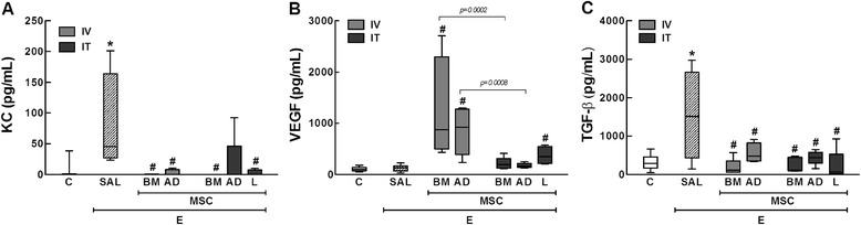
We sought to assess whether the effects of mesenchymal stromal cells (MSC) on lung inflammation and remodeling in experimental emphysema would differ according to MSC source and administration route. Emphysema was induced in C57BL/6 mice by intratracheal (IT) administration of porcine pancreatic elastase (0.1 UI) weekly for 1 month. After the last elastase instillation, saline or MSCs (1×10⁵), isolated from either mouse bone marrow (BM), adipose tissue (AD) or lung tissue (L), were administered intravenously (IV) or IT. After 1 week, mice were euthanized. Regardless of administration route, MSCs from each source yielded: 1) decreased mean linear intercept, neutrophil infiltration, and cell apoptosis; 2) increased elastic fiber content; 3) reduced alveolar epithelial and endothelial cell damage; and 4) decreased keratinocyte-derived chemokine (KC, a mouse analog of interleukin-8) and transforming growth factor-β levels in lung tissue. In contrast with IV, IT MSC administration further reduced alveolar hyperinflation (BM-MSC) and collagen fiber content (BM-MSC and L-MSC). Intravenous administration of BM- and AD-MSCs reduced the number of M1 macrophages and pulmonary hypertension on echocardiography, while increasing vascular endothelial growth factor. Only BM-MSCs (IV > IT) increased the number of M2 macrophages. In conclusion, different MSC sources and administration routes variably reduced elastase-induced lung damage, but IV administration of BM-MSCs resulted in better cardiovascular function and change of the macrophage phenotype from M1 to M2.


Figures

Figure 1
Figure 1
Schematic flow chart ( A ) and timeline ( B ) of the study design. C: intratracheal instillation of 50 μL of saline, E: intratracheal instillation of 0.1 UI of pancreatic porcine elastase (PPE), SAL: injection of 50 μL of saline, BM-MSC: bone marrow mesenchymal stromal cell (1 × 105) administration; AD-MSC: adipose tissue derived mesenchymal stromal cell (1 × 105) administration; L-MSC: lung derived mesenchymal stromal cell (1 × 105) administration; IV/IT: intravenous or intratracheal injection 3 h after the last instillation of saline or PPE; ♦: saline or PPE instillation; ★: all data were analyzed at day 28.
Figure 2
Figure 2
Representative photomicrographs of the lung parenchyma. C: control groups. E: emphysema groups. Mice were treated with saline (SAL) or bone marrow (BM), adipose (AD), and lung (L)-derived mesenchymal stromal cells (MSC). IV: intravenous route. IT: intratracheal route.
Figure 3
Figure 3
Collagen fibers in small airways (Picrosirius-polarization method). C: control groups. E: emphysema groups. Mice were treated with saline (SAL) or bone marrow (BM), adipose (AD), and lung (L)-derived mesenchymal stromal cells (MSC). IV: intravenous route. IT: intratracheal route. Values are mean ± SD of 7–30 mice in each group. All values were computed in ten random, non-coincident fields per animal. *Vs. C group (p < 0.05). #Vs. E-SAL group (p < 0.05).
Figure 4
Figure 4
Elastic fibers in the alveolar septa (Weigert’s resorcin fuchsin method). C: control groups. E: emphysema groups. Mice were treated with saline (SAL) or bone marrow (BM), adipose (AD), and lung (L)-derived mesenchymal stromal cells (MSC). IV: intravenous route. IT: intratracheal route. Values are mean ± SD of 7–30 mice in each group. All values were computed in ten random, non-coincident fields per animal. *Vs. C group (p < 0.05). #Vs. E-SAL group (p < 0.05).
Figure 5
Figure 5
Electron microscopy of lung parenchyma. C: control groups. E: emphysema groups. Mice were treated with saline (SAL), or bone marrow (BM), adipose (AD) and lung (L)-derived mesenchymal stem cells (MSC). In the C group, the alveolar epithelium is formed by type II pneumocytes (PII). Alveolar septa (AS) and capillaries (CAP) are intact. In E-SAL group, the AS is ruptured with capillary loss (arrows) and shows fibroblasts (FIB) and increase in collagen fibers. The alveolar epithelium was apparently normal but had zones with AS thickness containing no capillaries (arrows) even after intravenous or intratracheal BM-MSC and AD-MSC administration. After intratracheal L-MSC treatment, the AS is restored with new capillaries and collagen fibers are diminished.
Figure 6
Figure 6
Immunohistochemistry for iNOS ( A ) and arginase-1 ( B ). C: control groups. E: emphysema groups. Mice were treated with saline (SAL), or bone marrow (BM), adipose (AD) and lung (L)-derived mesenchymal stem cells (MSC). IV: intravenous route. IT: intratracheal route. Values are mean ± SD of 5–30 mice in each group. All values were computed in ten random, non-coincident fields per animal. *Vs. C group (p < 0.05). #Vs. E-SAL group (p < 0.05). Note positive cells in brown (arrow).
Figure 7
Figure 7
Levels of KC ( A ), VEGF ( B ), and TGF-β ( C ) in lung tissue. C: control groups. E: emphysema groups. Mice were treated with saline (SAL), or bone marrow (BM), adipose (AD) and lung (L)-derived mesenchymal stem cells (MSC). IV: intravenous route. IT: intratracheal route. Values are mean ± SD of 5–30 mice in each group. All values were computed in ten random, non-coincident fields per animal. *Vs. C group (p < 0.05). #Vs. E-SAL group (p < 0.05).
Figure 8
Figure 8
Short-axis B-dimensional views of both ventricles. The indices shown are ( A ) pulmonary artery acceleration time/pulmonary artery ejection time ratio – PAT/PET ratio – and ( B ) right ventricle area. (C) Echocardiographic images. LV: left ventricle, RV: right ventricle. The bars represent the means of 3 mice ± SD/group. C: control groups. E: emphysema groups. Mice were treated with saline (SAL), or bone marrow (BM), adipose (AD) and lung (L)-derived mesenchymal stem cells (MSC). IV: intravenous route. IT: intratracheal route. *Vs. C group (p < 0.05). #Vs. E-SAL group (p < 0.05).

References

    1. Minai OA, Benditt J, Martinez FJ. Natural history of emphysema. Proc Am Thorac Soc. 2008;5(4):468–474. doi: 10.1513/pats.200802-018ET. - DOI - PMC - PubMed
    1. From the Global Strategy for the Diagnosis, Management and Prevention of COPD, Global Initiative for Chronic Obstructive Lung Disease (GOLD).http://www.goldcopd.org/. - PubMed
    1. Weiss DJ. Concise review: current status of stem cells and regenerative medicine in lung biology and diseases. Stem Cells. 2014;32(1):16–25. doi: 10.1002/stem.1506. - DOI - PMC - PubMed
    1. Huh JW, Kim SY, Lee JH, Lee JS, Van Ta Q, Kim M, Oh YM, Lee YS, Lee SD. Bone marrow cells repair cigarette smoke-induced emphysema in rats. Am J Physiol Lung Cell Mol Physiol. 2011;301(3):L255–L266. doi: 10.1152/ajplung.00253.2010. - DOI - PubMed
    1. Kim SY, Lee JH, Kim HJ, Park MK, Huh JW, Ro JY, Oh YM, Lee SD, Lee YS. Mesenchymal stem cell-conditioned media recovers lung fibroblasts from cigarette smoke-induced damage. Am J Physiol Lung Cell Mol Physiol. 2012;302(9):L891–L908. doi: 10.1152/ajplung.00288.2011. - DOI - PubMed

Publication types

LinkOut - more resources