. 2014 Nov;3(11):1287-94.
doi: 10.5966/sctm.2014-0101. Epub 2014 Oct 1.

Cartilage repair using human embryonic stem cell-derived chondroprogenitors

Affiliations

Cartilage repair using human embryonic stem cell-derived chondroprogenitors

Aixin Cheng et al. Stem Cells Transl Med. 2014 Nov.

Abstract

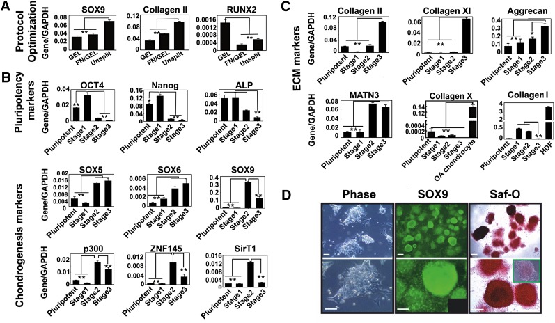
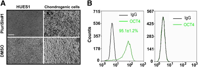
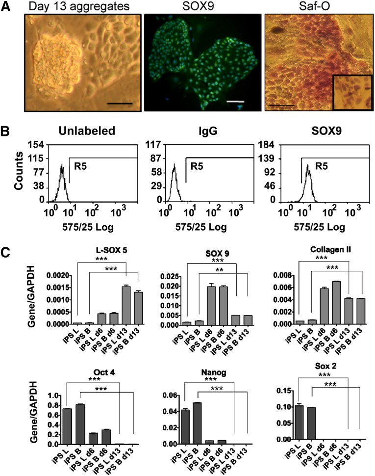
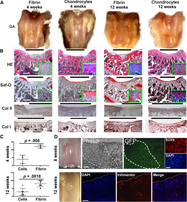
In initial work, we developed a 14-day culture protocol under potential GMP, chemically defined conditions to generate chondroprogenitors from human embryonic stem cells (hESCs). The present study was undertaken to investigate the cartilage repair capacity of these cells. The chondrogenic protocol was optimized and validated with gene expression profiling. The protocol was also applied successfully to two lines of induced pluripotent stem cells (iPSCs). Chondrogenic cells derived from hESCs were encapsulated in fibrin gel and implanted in osteochondral defects in the patella groove of nude rats, and cartilage repair was evaluated by histomorphology and immunocytochemistry. Genes associated with chondrogenesis were upregulated during the protocol, and pluripotency-related genes were downregulated. Aggregation of chondrogenic cells was accompanied by high expression of SOX9 and strong staining with Safranin O. Culture with PluriSln1 was lethal for hESCs but was tolerated by hESC chondrogenic cells, and no OCT4-positive cells were detected in hESC chondrogenic cells. iPSCs were also shown to generate chondroprogenitors in this protocol. Repaired tissue in the defect area implanted with hESC-derived chondrogenic cells was stained for collagen II with little collagen I, but negligible collagen II was observed in the fibrin-only controls. Viable human cells were detected in the repair tissue at 12 weeks. The results show that chondrogenic cells derived from hESCs, using a chemically defined culture system, when implanted in focal defects were able to promote cartilage repair. This is a first step in evaluating these cells for clinical application for the treatment of cartilage lesions.

Keywords: Arthritis; Cell transplantation; Embryonic stem cell; Tissue regeneration.


Figures

Figure 1.
Figure 1.
Characterization of cells subjected to the directed differentiation protocol. (A): The directed differentiation protocol was optimized by avoiding splitting the cells on day 12 and continuation of fibronectin substrate with gelatin to the end of the protocol, which resulted in a higher expression level of SOX9 and COL2A1, but lower RUNX2. The results are the means ± SD (n = 3). (B, C): Expression of genes associated with pluripotency and chondrogenesis (B) and chondrocyte ECM (C) during the directed the differentiation protocol was analyzed by quantitative reverse transcription polymerase chain reaction. Human dermal fibroblasts were used as a positive control for Collagen I. The results are the means ± SD (n = 3). Levels of significance are depicted as follows: ∗, p < .05; ∗∗, p < .01. (D): Chondrogenic cells derived from MAN7 showed a very high level of aggregation at the end of the protocol and high expression of SOX9 (inset shows IgG control), as well as a high level of Safranin O staining (inset shows pretreated with chondroitinase ABC). Scale bars = 100 μm. Abbreviations: ALP, alkaline phosphatase; ECM, extracellular matrix; FN, fibronectin; GAPDH, glyceraldehyde-3-phosphate dehydrogenase; GEL, gelatin; Saf-O, Safranin O.
Figure 2.
Figure 2.
Detection of residual pluripotent cells in human embryonic stem cell (hESC) chondrogenic cells. HUES1 and chondrogenic cells derived from these hESCs were treated with 2.5 μM PluriSIn1 or DMSO for 72 hours. (A): OCT4 expression in both types of cells was investigated with flow cytometry. (B): No significant difference was found for cells labeled with IgG control and OCT4. The results are the means ± SD (n = 3 experiments). Scale bar = 100 μm. Abbreviations: DMSO, dimethyl sulfoxide; NS, not significant.
Figure 3.
Figure 3.
Chondrogenic differentiation of iPSCs. iPSCs were differentiated into chondroprogenitors using the directed differentiation protocol. (A): At the end of the protocol, chondrogenic cell aggregation was observed, SOX9 expression was detected by immunofluorescence and the deposition of sulfated glycosaminoglycan was shown by Safranin O staining (inset shows cultures pretreated with chondroitinase ABC). Scale bars = 200 μm. (B): Flow cytometry study for SOX9 expression in chondrogenic cells from day 13 of the protocol. (C): Genes associated with pluripotency and chondrogenesis were measured by quantitative reverse transcription polymerase chain reaction. B and L indicate the two iPSC lines used for the experiment. The results are the means ± SD (n = 3). The levels of significance are depicted as follows: ∗∗, p < .01; ∗∗∗, p < .001. Abbreviations: d6, day 6; d13, day 13; GAPDH, glyceraldehyde-3-phosphate dehydrogenase; iPS, induced pluripotent stem cells; Saf-O, Safranin O.
Figure 4.
Figure 4.
In vivo cartilage formation promoted by chondrogenic cells derived from human embryonic stem cells. (A): RNU rats were killed at designated time points postimplantation, and the patella groove was assessed by macroscopic examination of the gross appearance after fixation. (B): Histological assessment of sections through knee cartilage using hematoxylin and eosin and Saf-O staining and by immunohistochemical staining for collagen I and II. (C): Cartilage repair scoring using Pineda’s system, which has a score, range from 0 (best) to 14 (worst). The results are the means ± SD (n = 3 animals for the 4 weeks group and n = 6 for the 12 weeks group; 18 animals in total) (D): GFP-positive cells (which expressed SOX9 before implantation, top right column) were detected 8 weeks after implantation of GFP-labeled chondrogenic cells (defect area outlined by dotted line). Human cells were detected 12 weeks after implantation by immunohistochemistry using anti-human vimentin antibody (surface of defect area shown by dotted line). Black bars = defect area; white bars = 500 μm; green bars = 100 μm. Abbreviations: Col, collagen; DAPI, 4′,6-diamidino-2-phenylindole; GA, gross appearance after fixation; GFP, green fluorescent protein; HE, hematoxylin and eosin; hVimentin, human vimentin; Saf-O, Safranin O.

References

    1. Brittberg M, Lindahl A, Nilsson A, et al. Treatment of deep cartilage defects in the knee with autologous chondrocyte transplantation. N Engl J Med. 1994;331:889–895. - PubMed
    1. Filardo G, Kon E, Berruto M, et al. Arthroscopic second generation autologous chondrocytes implantation associated with bone grafting for the treatment of knee osteochondritis dissecans: Results at 6 years. Knee. 2012;19:658–663. - PubMed
    1. Kang SW, Yoo SP, Kim BS. Effect of chondrocyte passage number on histological aspects of tissue-engineered cartilage. Biomed Mater Eng. 2007;17:269–276. - PubMed
    1. Hettrich CM, Crawford D, Rodeo SA. Cartilage repair: Third-generation cell-based technologies: Basic science, surgical techniques, clinical outcomes. Sports Med Arthrosc. 2008;16:230–235. - PubMed
    1. Pelttari K, Winter A, Steck E, et al. Premature induction of hypertrophy during in vitro chondrogenesis of human mesenchymal stem cells correlates with calcification and vascular invasion after ectopic transplantation in SCID mice. Arthritis Rheum. 2006;54:3254–3266. - PubMed

Publication types