. 2014 Dec 1;25(24):3926-41.
doi: 10.1091/mbc.E14-02-0708. Epub 2014 Oct 1.

Acute increase of α-synuclein inhibits synaptic vesicle recycling evoked during intense stimulation

Affiliations

Acute increase of α-synuclein inhibits synaptic vesicle recycling evoked during intense stimulation

David J Busch et al. Mol Biol Cell. .

Abstract

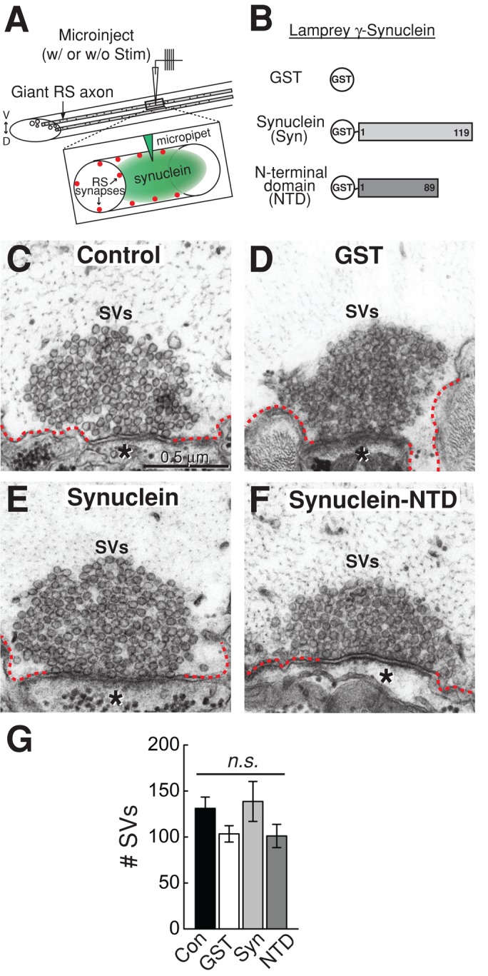
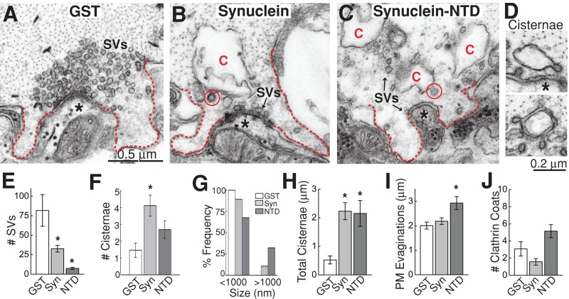
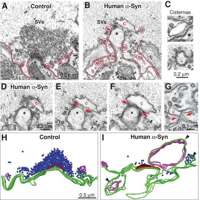
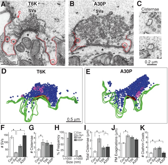
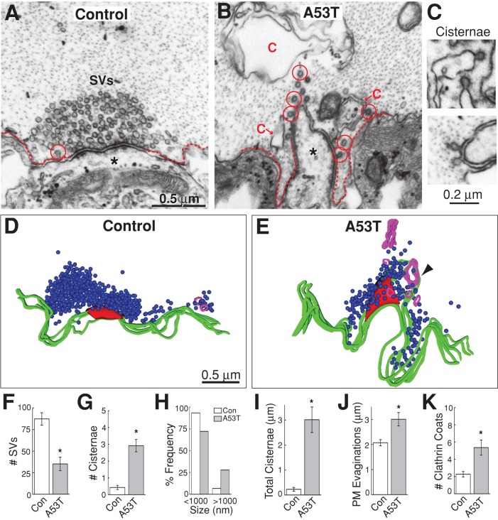
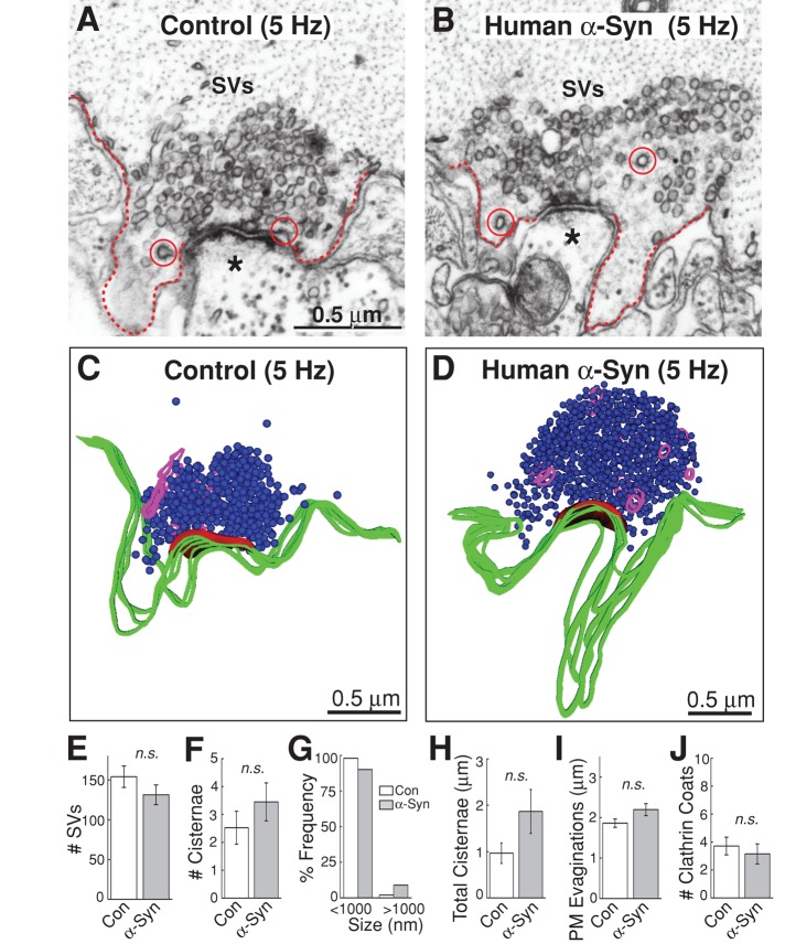
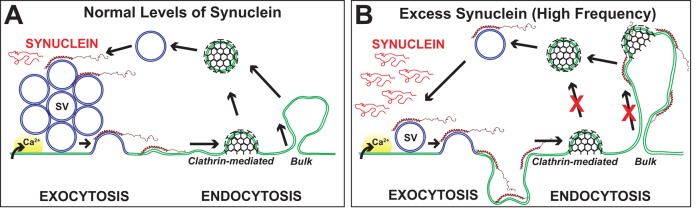
Parkinson's disease is associated with multiplication of the α-synuclein gene and abnormal accumulation of the protein. In animal models, α-synuclein overexpression broadly impairs synaptic vesicle trafficking. However, the exact steps of the vesicle trafficking pathway affected by excess α-synuclein and the underlying molecular mechanisms remain unknown. Therefore we acutely increased synuclein levels at a vertebrate synapse and performed a detailed ultrastructural analysis of the effects on presynaptic membranes. At stimulated synapses (20 Hz), excess synuclein caused a loss of synaptic vesicles and an expansion of the plasma membrane, indicating an impairment of vesicle recycling. The N-terminal domain (NTD) of synuclein, which folds into an α-helix, was sufficient to reproduce these effects. In contrast, α-synuclein mutants with a disrupted N-terminal α-helix (T6K and A30P) had little effect under identical conditions. Further supporting this model, another α-synuclein mutant (A53T) with a properly folded NTD phenocopied the synaptic vesicle recycling defects observed with wild type. Interestingly, the vesicle recycling defects were not observed when the stimulation frequency was reduced (5 Hz). Thus excess α-synuclein impairs synaptic vesicle recycling evoked during intense stimulation via a mechanism that requires a properly folded N-terminal α-helix.


Figures

FIGURE 1:
FIGURE 1:
Lamprey γ-synuclein is structurally similar to human α-synuclein. (A) Multiple sequence alignment of human synucleins (α, β, γ) and lamprey γ-synuclein. Black highlighted residues are those that are identical to human α-synuclein. Note the higher degree of similarity within the NTDs of all synucleins (aa 1–90; backlit in gray). Black lines indicate the 11-amino-acid repeats that characterize all synucleins and that fold into an α-helix. Epitope of the pan-synuclein antibody used in this study is indicated. (B) Top, solution NMR structure of human α-synuclein bound to detergent micelles (Ulmer et al., 2005); bottom, the predicted structure of lamprey γ-synuclein. Ribbon models were generated using University of California–San Francisco Chimera modeling software. (C and D) A pan-synuclein antibody generated against human α-synuclein recognizes all three recombinant human synucleins and GST-tagged lamprey γ-synuclein, indicating structural conservation. The antibody does not recognize GST alone. Top panels are the Western blots against synuclein (WB:Syn); bottom panels are Coomassie-stained (Coom) gels of the same proteins. (E and F) In rat brain lysates and in lamprey spinal cord (SC) and brain lysates, the pan-synuclein antibody recognizes an endogenous 17-kDa protein—the expected size for monomeric synuclein. (G) GST-tagged lamprey γ-synuclein and its NTD (Nterm) bind to suvs containing PC and PA in a 3:1 M ratio, but not to PC alone (n = 2). Neither the C-terminal domain (Cterm) of lamprey synuclein nor GST alone binds PC/PA, but instead remain as free protein. No binding to multilamellar vesicles (mlv) was observed.
FIGURE 2:
FIGURE 2:
Synuclein is localized to synaptic vesicle clusters at lamprey giant RS synapses. (A) Top, cross-section of a lamprey spinal cord stained with toluidine blue. Note the giant RS axons in the ventromedial tract. The box marks a single giant RS axon and the view shown in B–D. D, dorsal; V, ventral. Bottom, insets show the location of giant RS synapses along the periphery of the giant axons. SVs, synaptic vesicles. Asterisk marks postsynaptic dendrite. (B–G) Immunolabeling of synuclein (green) and SV2 (red) in the lamprey spinal cord. Merged image reveals strong colocalization between synuclein and SV2, indicating synuclein's localization at synaptic vesicle clusters (Pearson's colocalization coefficient = 0.78 ± 0.04; n = 5). Dotted line indicates the border of the giant RS axon. Arrows indicate RS synapses, which are shown in high magnification in panels E–G. Puncta outside the dotted line are synapses formed by smaller intraspinal axons within the neuropil. Scale bar in B is 20 μm and applies to panels B–D. Scale bar in E is 5 μm and applies to panels E–G.
FIGURE 3:
FIGURE 3:
Excess synuclein does not affect synaptic vesicle clustering at unstimulated synapses. (A) Experimental paradigm used in this study. Giant RS axons were microinjected with excess recombinant synuclein. This was followed by immediate fixation either without stimulation or after stimulation with action potentials. Inset shows how the proteins gain access to the presynaptic vesicle clusters. (B) Diagram of the recombinant proteins that were injected. (C–F) Electron micrographs of unstimulated giant RS synapses after no injection (Control) or after injection of GST (GST), full-length lamprey γ-synuclein (Synuclein), or its NTD (Synuclein-NTD). In all cases, the synaptic vesicle (SV) clusters are large and appear normal. Asterisks mark the postsynaptic dendrites. Dotted lines indicate the plasma membrane. Scale bar in C applies to panels C–F. (G) The number of SVs was not significantly different across any of these conditions. Bars represent mean ± SEM from n = 8–13 synapses, 2 axons. n.s., not significant by ANOVA.
FIGURE 4:
FIGURE 4:
Excess lamprey γ-synuclein inhibits synaptic vesicle recycling during intense stimulation. (A–C) Electron micrographs of stimulated (20 Hz, 5 min) RS synapses after treatment with GST alone, full-length γ-synuclein, or the NTD. In the GST controls, large synaptic vesicle (SV) clusters were visible, and plasma membrane extensions (dotted lines) were modest, owing to efficient SV recycling. In contrast, after treatment with synuclein or NTD, the number of vesicles was greatly reduced, and large cisternae (C) were apparent, indicating a defect in SV recycling. Cisternae often had clathrin-coated pits emanating from them (circles), suggesting they derived from the plasma membrane. Asterisks indicate postsynaptic dendrites. Scale bar in panel A applies to A–C. (D) Examples of cisternae after treatment with excess lamprey γ-synuclein. (E–J) Quantification of the endocytic defects produced by lamprey synuclein. Synuclein reduced the number of SVs at synapses (E) and increased the amount of membrane trapped in cisternae (F–H). Synuclein NTD additionally increased the size of plasma membrane evaginations (I). Changes in clathrin coats were not significant (J). Bars represent mean ± SEM from n = 13–35 synapses, 2–3 axons. Asterisks indicate statistical significance from GST controls by ANOVA (Tukey's post hoc p < 0.05).
FIGURE 5:
FIGURE 5:
Cisternae caused by excess lamprey γ-synuclein are often connected to the plasma membrane. (A–D) Serial electron micrographs through a stimulated RS synapse treated with lamprey γ-synuclein. In A, a single cisterna (C) appears to be disconnected from the plasma membrane. However, in B, the cisterna is contiguous with the plasma membrane (arrows), indicating that it is an extension of the plasma membrane. In C and D, the cisterna is no longer distinguishable from the plasmalemma. SVs, synaptic vesicles. Asterisks mark the postsynaptic dendrite. Scale bar in panel A applies to A–D. (E) Left, a large cisterna that has several clathrin-coated pits budding from it is shown, along with its connection to the plasma membrane; right, inset shows the neck of the bulk endocytic structure (arrows).
FIGURE 6:
FIGURE 6:
Excess human α-synuclein also inhibits synaptic vesicle recycling during intense stimulation. (A and B) Electron micrographs of stimulated RS synapses (20 Hz, 5 min) treated with buffer only (Control) or wild-type human α-synuclein. At control synapses, large synaptic vesicle (SV) clusters were visible, and plasma membrane evaginations (dotted lines) were modest because of efficient synaptic vesicle recycling. In contrast, after treatment of synapses with human α-synuclein, there were fewer SVs, cisternae were apparent, and plasma membrane evaginations were greatly expanded. Asterisks mark postsynaptic dendrites. C, cisterna. Circles, clathrin-coated pits or vesicles. Scale bar in A applies to B. (C) Cisternae caused by excess human α-synuclein. (D–G) Electron micrographs showing the connections between the cisternae and plasma membrane (arrows). (H and I) Three-dimensional reconstructions of stimulated RS synapses treated with buffer (control) or with excess human α-synuclein. The control synapse exhibits a large cluster of synaptic vesicles (blue) at the active zone (red), a relatively flat presynaptic plasma membrane (green), and small, infrequent cisternae (magenta). In contrast, after excess human α-synuclein, there were only a few synaptic vesicles, very large plasma membrane evaginations, and several larger, atypical cisternae. Most large cisternae were connected to the plasma membrane in at least one section (arrowheads). Scale bar in H applies to I.
FIGURE 7:
FIGURE 7:
α-Synuclein mutations A30P and T6K alter proper folding of the N-terminal α-helix. (A) Wild-type (WT) human α-synuclein and mutants. A30P and T6K exhibit significantly reduced α-helical content in the NTD, based on circular dichroism analysis (Perrin et al., 2000). Asterisks mark the locations of the mutations. (B) Liposome flotation assay. Liposomes (circles), and thus bound protein (bars), are predominantly in the top two fractions after centrifugation, while unbound protein is in the lower fractions, where the percentage of liposomes is low. The data in the graph show an example of fluorescence measurements from NBD-labeled PC liposomes. (C) WT α-synuclein binds to PC/PA liposomes but not to PC alone. T6K and A30P also bind to PC/PA liposomes, but with reduced affinity, confirming previous observations (Perrin et al., 2000). (D) Quantification of data from liposome-binding assays showing α-synuclein and mutants binding to PC and PC/PA. Data points represent mean ± SEM from n = 3 independent experiments.
FIGURE 8:
FIGURE 8:
Endocytic defects caused by excess human α-synuclein require proper folding of the N-terminal α-helix. (A and B) T6K and A30P greatly reduced the synaptic vesicle recycling defects after 20-Hz, 5-min stimulation. Synaptic vesicle (SV) clusters were larger, and the cisternae were less prevalent than with WT α-synuclein (see Figure 6). Asterisks mark the postsynaptic dendrites. Dotted lines indicate the plasma membrane. C, cisterna; circles, clathrin-coated pits or vesicles. Scale bar in A applies to B. (C) The cisternae observed with excess T6K (top) or A30P (bottom) were smaller and discontinuous with the plasma membrane (see D and E). (D and E) Three-dimensional reconstructions of stimulated RS synapses treated with T6K and A30P do not exhibit major endocytic defects but instead appear more similar to controls (compare with Figure 6H). Scale bar in D applies to E. (F–K) Quantification of the ultrastructural analyses. Consistent with an inhibition in synaptic vesicle recycling, wild-type α-synuclein decreased the number of synaptic vesicles (F), while increasing the number of cisternae (G), the frequency of large cisternae (H), the amount of membrane trapped within cisternae (I), and the size of plasma membrane evaginations (J). The increase in clathrin-coated pits and vesicles with α-synuclein also indicates vesicle recycling defects (K). In contrast, the T6K and A30P mutations reduced all aspects of the vesicle recycling defects associated with α-synuclein. Bars represent mean ± SEM from n = 22–44 synapses, 2–4 axons. Asterisks indicate statistical significance by ANOVA (Tukey's post hoc p < 0.05).
FIGURE 9:
FIGURE 9:
A53T also impairs synaptic vesicle recycling evoked by high-frequency stimulation. (A and B) Compared with control synapses, A53T caused a reduction in the number of synaptic vesicles (SVs) and expansion of the plasma membrane (dotted lines) after 20-Hz, 5-min stimulation. Large cisternae were also apparent. C, cisterna; circles, clathrin-coated pits or vesicles. Scale bar in A applies to B. (C) Cisternae caused by A53T. (D and E) Three-dimensional reconstructions show the endocytic phenotype caused by A53T. A large cisterna was connected to the plasma membrane (arrowhead). Scale bar in D applies to E. (F–K) Synapses treated with A53T induced the same phenotype as wild-type α-synuclein: a reduction in synaptic vesicles (F); an increase in the number, size, and amount of membrane within cisternae (G–I); an increase in plasma membrane evaginations (J); and an increase in the number of clathrin-coated pits and vesicles (K). Bars represent mean ± SEM from n = 25–35 synapses, 2 axons. Asterisks indicate statistical significance by Student's t test (p < 0.05).
FIGURE 10:
FIGURE 10:
α-Synuclein does not affect synaptic vesicle recycling evoked by lower frequency stimulation. (A and B) Following a lower-frequency stimulation (5 Hz, 30 min), synapses treated with excess human α-synuclein appeared similar to control synapses. No endocytic phenotype was observed. Dotted lines indicate plasma membrane. SVs, synaptic vesicles; circles, clathrin-coated pits or vesicles. Asterisks mark the postsynaptic dendrites. Scale bar in A applies to B. (C and D) Three-dimensional reconstructions of control and α-synuclein treated synapses after 5-Hz, 30-min stimulation. No obvious differences were observed. (E–J) Quantification revealed that there was no significant change in the number of synaptic vesicles (E); the number, size distribution, or amount of membrane within cisternae (F–H); the size of plasma membrane evaginations (I); or the number of clathrin coats (J). Bars represent mean ± SEM from n = 20–22 synapses, 2 axons. n.s., not significant by Student's t test (p > 0.05).
FIGURE 11:
FIGURE 11:
Model for how excess α-synuclein impairs synaptic vesicle recycling. (A) Synaptic vesicle (SV) recycling with endogenous levels of synuclein. After calcium (Ca2+) influx and exocytosis, vesicles are efficiently recycled via local endocytosis from the plasma membrane. (B) With excess α-synuclein, SV recycling is disrupted, as evidenced by a loss of SVs and an expansion of the plasma membrane, which often buckled inward to create apparent cisternae. Excess α-synuclein inhibited at least two modes of synaptic vesicle recycling, clathrin-mediated and bulk endocytosis, but only during high-frequency stimulation. The working model is that excess α-synuclein, in its α-helical conformation, masks or mislocalizes key lipids or proteins necessary for initiating synaptic vesicle recycling.

References

    1. Abeliovich A, Schmitz Y, Farinas I, Choi-Lundberg D, Ho WH, Castillo PE, Shinsky N, Verdugo JM, Armanini M, Ryan A, et al. Mice lacking alpha-synuclein display functional deficits in the nigrostriatal dopamine system. Neuron. 2000;25:239–252. - PubMed
    1. Andersson F, Jakobsson J, Low P, Shupliakov O, Brodin L. Perturbation of syndapin/PACSIN impairs synaptic vesicle recycling evoked by intense stimulation. J Neurosci. 2008;28:3925–3933. - PMC - PubMed
    1. Bendor JT, Logan TP, Edwards RH. The function of alpha-synuclein. Neuron. 2013;79:1044–1066. - PMC - PubMed
    1. Boassa D, Berlanga ML, Yang MA, Terada M, Hu J, Bushong EA, Hwang M, Masliah E, George JM, Ellisman MH. Mapping the subcellular distribution of alpha-synuclein in neurons using genetically encoded probes for correlated light and electron microscopy: implications for Parkinson's disease pathogenesis. J Neurosci. 2013;33:2605–2615. - PMC - PubMed
    1. Bolte S, Cordelieres FP. A guided tour into subcellular colocalization analysis in light microscopy. J Microsc. 2006;224:213–232. - PubMed

Publication types

MeSH terms

LinkOut - more resources