

Longitudinal assessment of concurrent changes in left ventricular ejection fraction and left ventricular myocardial tissue characteristics after administration of cardiotoxic chemotherapies using T1-weighted and T2-weighted cardiovascular magnetic resonance
- PMID: 25273568
- PMCID: PMC4241241
- DOI: 10.1161/CIRCIMAGING.114.002217
Longitudinal assessment of concurrent changes in left ventricular ejection fraction and left ventricular myocardial tissue characteristics after administration of cardiotoxic chemotherapies using T1-weighted and T2-weighted cardiovascular magnetic resonance
Abstract
Background: In a murine anthracycline-related cardiotoxicity model, increases in cardiovascular magnetic resonance myocardial contrast-enhanced T1-weighted signal intensity are associated with myocellular injury and decreases with left ventricular ejection fraction. We sought to determine whether T1- and T2-weighted measures of signal intensity associate with decreases in left ventricular ejection fraction in human subjects receiving potentially cardiotoxic chemotherapy.
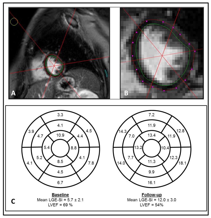
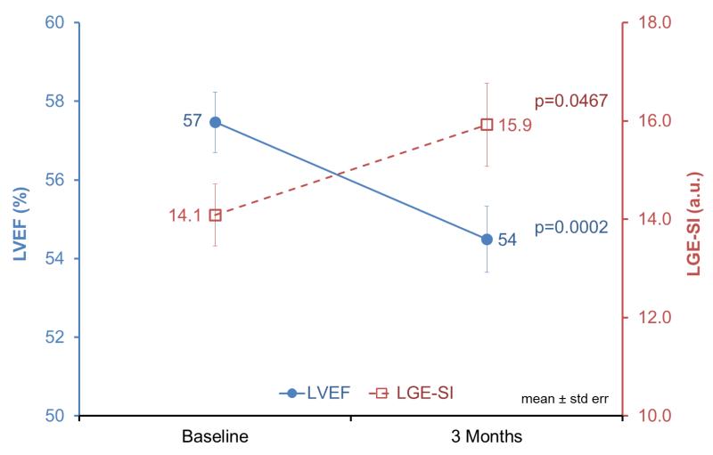
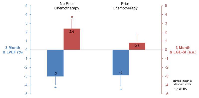
Methods and results: In 65 individuals with breast cancer (n=51) or a hematologic malignancy (n=14), we measured left ventricular volumes, ejection fraction, and contrast-enhanced T1-weighted and T2-weighted signal intensity before and 3 months after initiating potentially cardiotoxic chemotherapy using blinded, unpaired analysis of cardiovascular magnetic resonance images. Participants were aged 51 ± 12 years, of whom 55% received an anthracycline, 38% received a monoclonal antibody, and 6% received an antimicrotubule agent. Overall, left ventricular ejection fraction decreased from 57 ± 6% to 54 ± 7% (P<0.001) because of an increase in end-systolic volume (P<0.05). T1-weighted signal intensities also increased from 14.1 ± 5.1 to 15.9 ± 6.8 (P<0.05), with baseline values trending higher among individuals who received chemotherapy before study enrollment (P=0.06). Changes in T1-weighted signal intensity did not differ within the 17 LV myocardial segments (P=0.97). Myocardial edema quantified from T2-weighted images did not change significantly after 3 months (P=0.70).
Conclusions: Concordant with previous animal studies, cardiovascular magnetic resonance measures of contrast-enhanced T1-weighted signal intensity occur commensurate with small but significant left ventricular ejection fraction declines 3 months after the receipt of potentially cardiotoxic chemotherapy. These data indicate that changes in T1-weighted signal intensity may serve as an early marker of subclinical injury related to the administration of potentially cardiotoxic chemotherapy in human subjects.
Keywords: anthracyclines; cardiotoxicity; chemotherapy; left ventricular function; magnetic resonance imaging.
© 2014 American Heart Association, Inc.
Figures
References
-
- Ewer MS, Yeh E. Cancer and the heart. BC Decker Inc.; Lewiston, NY: 2006. Print.
-
- Wassmuth R, Lentzsch S, Erdbruegger U, Schulz-Menger J, Doerken B, Dietz R, Friedrich MG. Subclinical cardiotoxic effects of anthracyclines as assessed by magnetic resonance imaging- A pilot study. Am Heart J. 2001;141:1007–1013. - PubMed
-
- Thompson RC, Canby RC, Lojeski EW, Ratner AV, Fallon JT, Pohost GM. Adriamycin cardiotoxicity and proton nuclear magnetic resonance relaxation properties. Am Heart J. 1987;113:1444–1449. - PubMed
-
- Cottin Y, Ribuot C, Maupoil V, Godin D, Arnould L, Brunotte F, Rochette L. Early incidence of adriamycin treatment on cardiac parameters in the rat. Can J Physiol Pharmacol. 1994;72:140–145. - PubMed
Publication types
MeSH terms
Substances
Grants and funding
LinkOut - more resources
Full Text Sources
Other Literature Sources
Medical
