. 2014 Oct 2;16(5):461.
doi: 10.1186/s13075-014-0461-9.

Activation of fibroblast-like synoviocytes derived from rheumatoid arthritis via lysophosphatidic acid-lysophosphatidic acid receptor 1 cascade

Activation of fibroblast-like synoviocytes derived from rheumatoid arthritis via lysophosphatidic acid-lysophosphatidic acid receptor 1 cascade

Yoshishige Miyabe et al. Arthritis Res Ther. .

Abstract

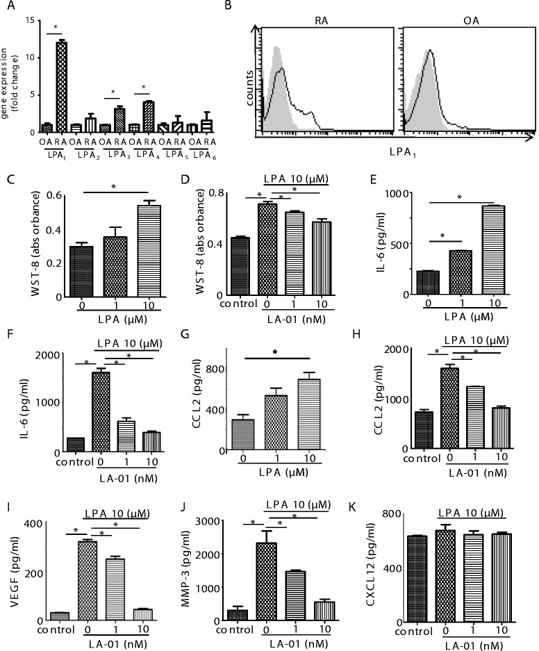
Introduction: Lysophosphatidic acid (LPA) is a bioactive lipid that binds to G protein-coupled receptors (LPA1-6). Recently, we reported that abrogation of LPA receptor 1 (LPA1) ameliorated murine collagen-induced arthritis, probably via inhibition of inflammatory cell migration, Th17 differentiation and osteoclastogenesis. In this study, we examined the importance of the LPA-LPA1 axis in cell proliferation, cytokine/chemokine production and lymphocyte transmigration in fibroblast-like synoviocytes (FLSs) obtained from the synovial tissues of rheumatoid arthritis (RA) patients.

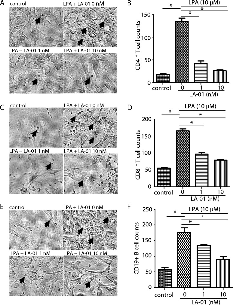
Methods: FLSs were prepared from synovial tissues of RA patients. Expression of LPA1-6 was examined by quantitative real-time RT-PCR. Cell surface LPA1 expression was analyzed by flow cytometry. Cell proliferation was analyzed using a cell-counting kit. Production of interleukin 6 (IL-6), vascular endothelial growth factor (VEGF), chemokine (C-C motif) ligand 2 (CCL2), metalloproteinase 3 (MMP-3) and chemokine (C-X-C motif) ligand 12 (CXCL12) was measured by enzyme-linked immunosorbent assay. Pseudoemperipolesis was evaluated using a coculture of RA FLSs and T or B cells. Cell motility was examined by scrape motility assay. Expression of adhesion molecules was determined by flow cytometry.

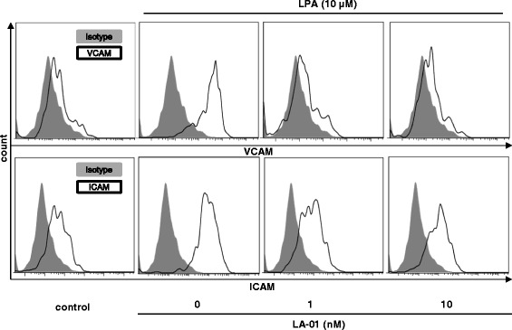
Results: The expression of LPA1 mRNA and cell surface LPA1 was higher in RA FLSs than in FLSs from osteoarthritis tissue. Stimulation with LPA enhanced the proliferation of RA FLSs and the production of IL-6, VEGF, CCL2 and MMP-3 by FLSs, which were suppressed by an LPA1 inhibitor (LA-01). Ki16425, another LPA1 antagonist, also suppressed IL-6 production by LPA-stimulated RA FLSs. However, the production of CXCL12 was not altered by stimulation with LPA. LPA induced the pseudoemperipolesis of T and B cells cocultured with RA FLSs, which was suppressed by LPA1 inhibition. In addition, LPA enhanced the migration of RA FLSs and expression of vascular cell adhesion molecule and intercellular adhesion molecule on RA FLSs, which were also inhibited by an LPA1 antagonist.

Conclusions: Collectively, these results indicate that LPA-LPA1 signaling contributes to the activation of RA FLSs.


Figures

Figure 1
Figure 1
Expression of lysophosphatidic acid receptors and the effect of lysophosphatidic acid receptor 1 on proliferation and production of inflammatory mediators in rheumatoid arthritis fibroblast-like synoviocytes. The expression levels of lysophosphatidic acid receptor 1 through 6 (LPA1–6) mRNA in fibroblast-like synoviocytes (FLSs) derived from the rheumatoid arthritis (RA) synovium (n = 10) were compared to those in FLSs from osteoarthritis (OA) synovium (n = 5) by real-time RT-PCR (A). Data were derived from samples from multiple individuals. Data are presented as the mean ± SEM. *P < 0.05 for RA vs OA. Cell surface expression of LPA1 on RA (n = 5) and OA (n = 3) FLSs was analyzed by flow cytometry (B). Filled histogram (gray): isotype control; open histogram (black line): LPA1. Representative histograms are shown. RA FLSs were cultured with lysophosphatidic acid (LPA) for 72 hours (C). FLSs were preincubated with an LPA1 inhibitor, LA-01, for 30 minutes,then stimulated with 10 μM LPA for 72 hours (D). Control: no stimulation with LPA. Cell proliferation was measured by using a cell counting kit (C) and (D). RA FLSs were cultured with LPA for 24 hours. Concentrations of interleukin 6 (IL-6) and chemokine (C-C motif) ligand 2 (CCL2) in the culture supernatant were measured by enzyme-linked immunosorbent assay (ELISA) (E) and (G). FLSs were preincubated with LA-01 for 30 minutes, then stimulated with 10 μM LPA for 24 hours. Concentrations of IL-6, CCL2, vascular endothelial growth factor (VEGF), matrixmetalloproteinase (MMP-3) and CXCL12 in the culture supernatant were measured by ELISA (F), and (H) through (K). Control: no stimulation with LPA. Data are presented as the means (±SEM) of one of three independent experiments analyzed in triplicate. *P < 0.05 vs control or LA-01 0 nM (C) through (K).
Figure 2
Figure 2
Effect of lysophosphatidic acid receptor 1 on pseudoemperipolesis and migration of rheumatoid arthritis fibroblast-like synoviocytes. After preincubation of cocultured rheumatoid arthritis (RA) fibroblast-like synoviocytes (FLSs) and CD4+ T cells (A) and (B) or CD8+ T cells (C) and (D) or CD19+ B cells (E) and (F) with a lysophosphatidic acid (LPA) receptor 1 inhibitor (LA-01; 0, 1 or 10 nM) for 30 minutes, the cells were stimulated with 10 μM LPA for 12 hours. Control: no stimulation with LPA. After the cells were washed, the number of lymphocytes beneath FLSs was counted. Representative photomicrographs of three independent experiments are shown (A, C and E). Arrows indicate the lymphocytes beneath FLSs. Original magnification, ×200. Data on the number of lymphocytes beneath FLSs are presented as one of three independent experiments analyzed in triplicate (B, D, and F). Data are presented as the mean ± SEM. *P < 0.05 vs control or LA-01 0 nM (B, D, F).
Figure 3
Figure 3
The effect of lysophosphatidic acid receptor 1 on the migration of rheumatoid arthritis fibroblast-like synoviocytes. A scraped cell-free area was created on cultured RA FLSs. After preincubation with a lysophosphatidic acid (LPA) receptor 1 inhibitor (LA-01; 0, 1 or 10 nM) for 30 minutes, cells were stimulated with 10 μM LPA for 48 hours. Control: no stimulation with LPA. (A) Representative photomicrographs of three independent experiments are shown. Original magnification, ×40. (B) The cell-free area was assessed, and a ratio (cell-free area in 48 hours to cell-free area in 0 hours) was defined. Data are presented as the means (±SEM) of one of three independent experiments analyzed in triplicate. *P < 0.05 vs control or LA-01 0 nM. Upper dashed line indicates the cells are stimulated with LPA 10 uM, and lower dashed line indicates LA-01 is added with indicated concentration.
Figure 4
Figure 4
The effect of lysophosphatidic acid receptor 1 on the expression of adhesion molecules on fibroblast-like synoviocytes. Rheumatoid arthritis (RA) fibroblast-like synoviocytes (FLSs) were pretreated with a lysophosphatidic acid (LPA) receptor 1 inhibitor (LA-01; 0, 1 or 10 nM) for 30 minutes, then the cells were stimulated with 10 μM LPA for 12 hours. Cells were stained with allophycocyanin-conjugated monoclonal antibody (mAb) against vascular cell adhesion molecule (anti-VCAM) or phycoerythrin-conjugated mAb against intercellular adhesion molecule (anti-ICAM). Allophycocyanin- or phycoerythrin-conjugated mouse immunoglobulin G1 (IgG1) was used as a control. The expression of VCAM and ICAM on FLSs was analyzed by flow cytometry. Filled histogram (gray): isotype control; open histogram (black line): VCAM or ICAM.

Similar articles

Cited by

References

    1. Kinne RW, Bräuer R, Stuhlmüller B, Palombo-Kinne E, Burmester GR. Macrophages in rheumatoid arthritis. Arthritis Res. 2000;2:189–202. doi: 10.1186/ar86. - DOI - PMC - PubMed
    1. Bottini N, Firestein GS. Duality of fibroblast-like synoviocytes in RA: passive responders and imprinted aggressors. Nat Rev Rheumatol. 2013;9:24–33. doi: 10.1038/nrrheum.2012.190. - DOI - PMC - PubMed
    1. Aoki J. Mechanisms of lysophosphatidic acid production. Semin Cell Dev Biol. 2004;15:477–489. doi: 10.1016/j.semcdb.2004.05.001. - DOI - PubMed
    1. Kawagoe H, Stracke ML, Nakamura H, Sano K. Expression and transcriptional regulation of the PD-Iα/autotaxin gene in neuroblastoma. Cancer Res. 1997;57:2516–2521. - PubMed
    1. Yang SY, Lee J, Park CG, Kim S, Hong S, Chung HC, Min SK, Han JW, Lee HW, Lee HY. Expression of autotaxin (NPP-2) is closely linked to invasiveness of breast cancer cells. Clin Exp Metastasis. 2002;19:603–608. doi: 10.1023/A:1020950420196. - DOI - PubMed

Publication types

MeSH terms

Substances

LinkOut - more resources