. 2014 Oct 1:13:226.
doi: 10.1186/1476-4598-13-226.

Elevated autocrine EDIL3 protects hepatocellular carcinoma from anoikis through RGD-mediated integrin activation

Affiliations

Elevated autocrine EDIL3 protects hepatocellular carcinoma from anoikis through RGD-mediated integrin activation

Ming-Xuan Feng et al. Mol Cancer. .

Abstract

Background: A remolded microenvironment in hepatocellular carcinoma (HCC) caused by abnormally expressed matricellular proteins could promote HCC progression. The cell-matrix interactions mediated by integrins play an important role in tumor microenvironment. Epidermal Growth Factor-like repeats and Discoidin I-Like Domains 3 (EDIL3), an extracellular matrix (ECM) protein with angiogenic and anti-inflammatory effects, is abnormally highly expressed in HCC. Here we aim to analyze its expression in liver and HCC tissues, investigate the underlined mechanisms accounted for HCC progression.

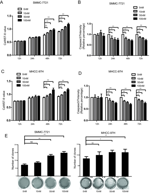
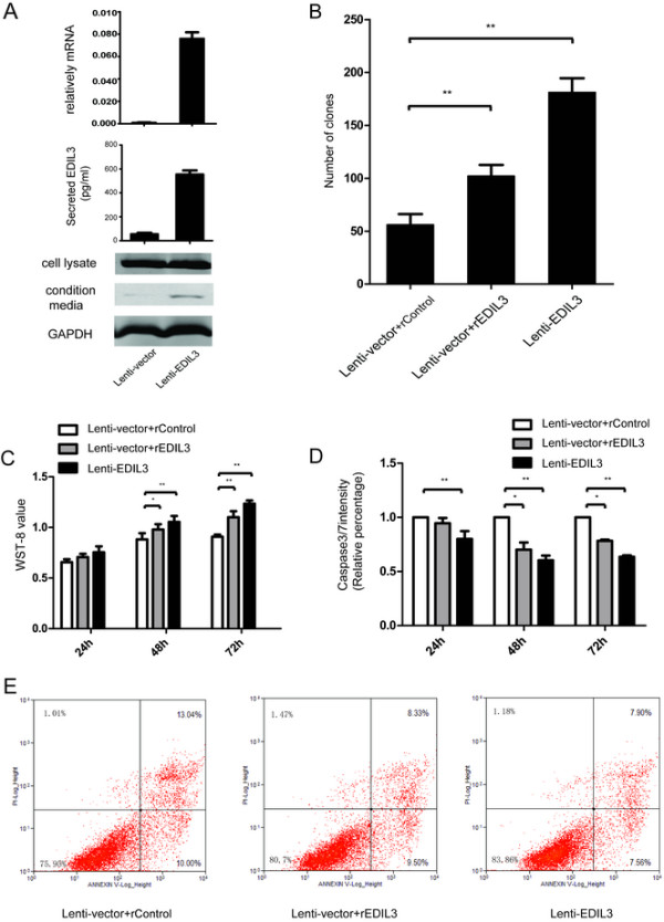
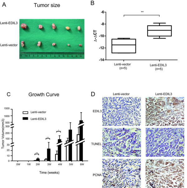
Methods: EDIL3 expression level is examined in normal liver, cirrhotic liver and HCC at both mRNA and protein level. The association between EDIL3 and clinical outcomes is analyzed. The pattern of EDIL3 expression and location is examined using Immunofluorescence and ELISA. Overexpression or knock-down of EDIL3 in a panel of cell lines are subjected to assays related to proliferation, invasion, and anoikis to investigate the mechanisms of this matrix protein in HCC progression. Recombinant EDIL3 treatment is applied to confirm the results.

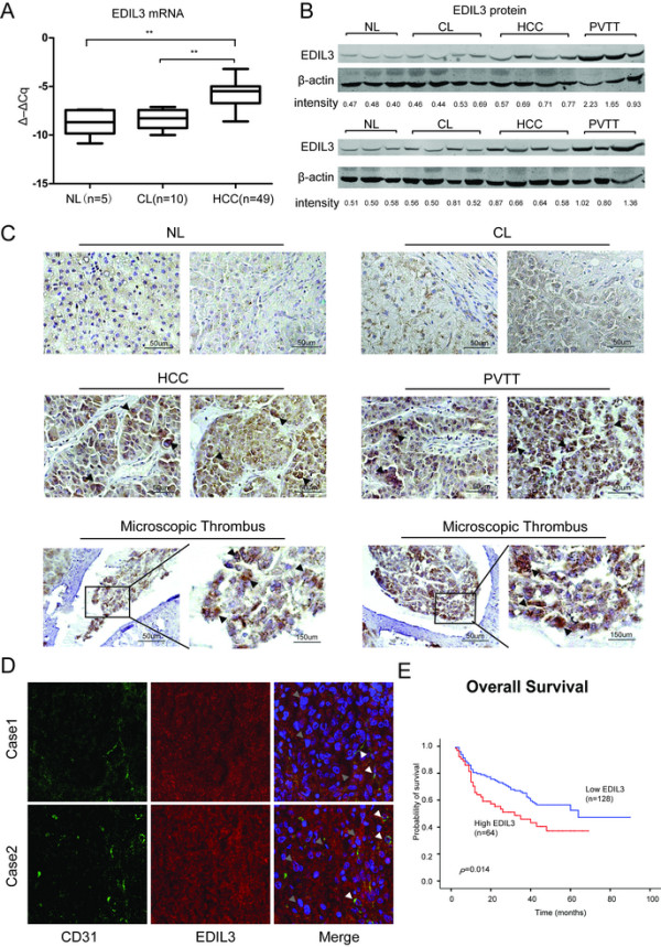
Results: Compared with normal liver and cirrhotic liver, EDIL3 is elevated in HCC. High level of EDIL3 protein is much more commonly in patients with larger tumor or portal vein tumor thrombus (PVTT) formation, associated with poor prognosis. EDIL3 is abundantly expressed in HCC cells and secreted by cancer cells. In vitro and in vivo studies indicate that EDIL3, probably in an autocrine manner, inhibits anoikis and promotes anchorage-independent growth of HCC cells. Further mechanistic studies suggest integrin ligation by EDIL3 and thus that the sustained activation of the FAK-Src-AKT signal is responsible for the anoikis resistance and anchorage independence. Both the administration of cilengitide, a RGD-containing integrin antagonist, and silencing of integrin αV, an important RGD-binding integrin, results in the blockade of anoikis-resistance induced by EDIL3.

Conclusion: Our study suggests that high levels of autocrine EDIL3 may contribute to a receptive microenvironment for the survival of detached HCC cells and may involve in cancer cell spreading. We also highlight the importance of interaction between EDIL3 and integrin αV and suggest disrupting the ligation of EDIL3 to integrins via RGD-blocking in selected patients may bear potential therapeutic value.


Figures

Figure 1
Figure 1
EDIL3 is significantly up-regulated in HCC and PVTT compared with normal liver (NL) and cirrhotic livers (CL) and is closely related to the prognosis of HCC. A, The transcriptional level of EDIL3 is measured via qRT-PCR in 5 NLs, 10 CLs and 49 HCCs. The relative mRNA level is normalized to β-actin and is presented as Δ–ΔCq. The mRNA of EDIL3 in HCC is significantly higher than both NL and CL; B, The protein level of EDIL3 in NL, CL, HCC and PVTT was examined by western blot with β-actin as a loading control. The EDIL3/β-actin densitometry is performed and shown as density value below. EDIL3 is mildly expressed In NL and CL while significantly elevated in HCC and PVTT; C, Representative pictures demonstrating EDIL3 staining in different liver samples including NL, CL, HCC, PVTT and microscopic thrombi by IHC. Scale bars, 50 μm or 150 μm; Arrow heads indicate that high EDIL3 expression is largely localized to cancer cells. D, Confocal microscopic observation of immunofluorescence staining of CD31 (green), an endothelium marker, and EDIL3 (red) in HCC samples show EDIL3 is not only localized with endothelium, but also widely and diffusively located in cancer cells clusters. White arrows indicates the endothelium; Grey arrows indicates the cancer cell cluster. E, Kaplan-Meier analysis of overall survival between EDIL3-negative or moderately positive patients and highly positive patients shows a significant survival advantage in EDIL3 low express group. **: P < 0.01.
Figure 2
Figure 2
EDIL3 exhibits a unique expression pattern in cell lines. A, detailed analysis of EDIL3 expression in mRNA level, cell lysates and CMs of 7 HCC and 2 non-HCC cell lines demonstrated an approximate correlation between mRNA and secreted EDIL3 in CMs, whereas EDIL3 in cell lysates exhibited almost same intensity. Notably, Huh-7 and CSQT-2 exhibited a much higher level of secreted EDIL3. B, ELISA assay testing the EDIL3 in CMs of 9 cell lines validates the secreted EDIL3 level is approximately correlated with mRNA level. C, Immunofluorescence staining of EDIL3, F-actin and DAPI in confocal microscope shows EDIL3 is localized within cells and at almost the same intensity in all the 9 cell lines under test, despite their varied mRNA level.
Figure 3
Figure 3
Treatment of EDIL3 contributes to anoikis resistance and anchorage-independent growth advantage in HCC. A, Different concentration of recombinant EDIL3 is used to treat SMMC-7721 suspended in poly-hema coated dishes. It sustains the viability of SMMC-7721 as demonstrated by caspase3WSt-8 assay in a time and dose dependent manner compared with control protein. B, Recombinant EDIL3 ameliorate the anoikis compared with control protein in SMMC-7721 as demonstrated by caspase3/7 intensity assay in a time and dose dependent manner. C-D, MHCC-97H is subjected to the assays same as SMMC-7721 and obtain consistent result. E, Administration of recombinant EDIL3 increases the anchorage-independent growth of SMMC-7721 and MHCC-97H cells in soft agar compared with control protein in a dose dependent manner. The assays last for 28 days. *: P < 0.05; **: P < 0.01.
Figure 4
Figure 4
Overexpression of EDIL3 in SMMC-7721 cells significantly increases the anoikis resistance and anchorage independence. A, Overexpression of EDIL3 is validated by real-time PCR, western blot and ELISA assay, confirming that the increase in transcription leads to a higher autocrine EDIL3. B, EDIL3-overexpressing SMMC-7721 forms more clones in soft agar compared with control cells, which can be partly mimicked by recombinant EDIL3 (50 nM). C, After being suspended for up to 72 h, the overexpressing group demonstrates a high WST-8 value compare to the control cells at 48 h and 72 h, and administration of recombinant EDIL3(50 nM) in control cells partly mimics the effect induced by EDIL3 overexpression. D, Caspase3/7 intensity assay validate that the difference in cell viability between the two groups is caused by lower apoptosis induction. E, Annexin V/PI stain by FACS is performed to validate the apoptosis result. EDIL3 overexpression lowers both the early phase and later phase apoptosis percentage and recombinant protein partly mimics this effect. *: P < 0.05; **: P < 0.01.
Figure 5
Figure 5
EDIL3 promotes HCC tumorigenesis in vivo. A total of 1.0 × 106 of EDIL3-overexpressing or control SMMC-7721 cells are subcutaneously implanted into the right flank of 5 nude mice of each groups. The mice were observed and tumors formed are measured every week and resected after 6 weeks. A, After 6 weeks, EDIL3-overexpressing group shows relatively larger tumors compare with control group. B, qRT-PCR of 5 tumors in each group demonstrates a significant elevation in mRNA level. C, Tumor growth curve reveals a shorter latency in the EDIL3-overexpressing group (2 weeks) vs. the control group (4 weeks) and tumors in the EDIL3-overexpressing group are significantly larger than control group from 2nd week to 4th week. D, IHC stain in the same region of the tumor confirms the overexpression of EDIL3 and suggests more active proliferation (PCNA) and lower apoptosis (tunel) upon EDIL3 overexpression. **: P < 0.01.
Figure 6
Figure 6
Sustained activation of FAK-Src by EDIL3 through RGD recognition. A, Western blot and densimetric analysis suggests EDIL3 overexpression sustains the signal intensity of FAK-Src and results in higher AKT phosphorylation within suspended SMMC-7721 cells over 48 hours. The elevated 397p-FAK, 416p-Src and 473p-AKT axis in overexpressing cells compare with control cells exists at most of the time points. B, Cilengitide (10 μM) reversed the activation of the FAK-Src-AKT axis induced by EDIL3. The signal intensity was examined after 24 h of suspension.
Figure 7
Figure 7
Disrupting integrin-EDIL3 ligation deprives HCC of anoikis resistance induced by EDIL3 in SMMC-7721 and MHCC-LM3. A, Cilengitide (10 μM) reduces the WST-8 value and increased casepase3/7 of the EDIL3 overexpressing group back to levels of control group, suggesting a blockage of EDIL3’s effect. There was not any effect observed in the control group in WST-8 and caspase3/7 assay. Both the two cell lines show consistent results. B, when integrin αV was knocked down to a very low level in both cell lines by two siRNAs (Si2 and Si3), the alteration in WST-8 and casepase3/7 value of EDIL3-overexpressing cells compared with control cells disappear. Interestingly, the WST-8 value and casepase3/7 intensity results suggest the overall anoikis significantly declined upon integrin αV silencing in both siRNA knock-down groups compare with si-control group. **: P < 0.01.

References

    1. Forner A, Llovet JM, Bruix J. Hepatocellular carcinoma. Lancet. 2012;379:1245–1255. doi: 10.1016/S0140-6736(11)61347-0. - DOI - PubMed
    1. Farazi PA, DePinho RA. Hepatocellular carcinoma pathogenesis: from genes to environment. Nat Rev Cancer. 2006;6:674–687. doi: 10.1038/nrc1934. - DOI - PubMed
    1. Yang JD, Nakamura I, Roberts LR. The tumor microenvironment in hepatocellular carcinoma: Current status and therapeutic targets. Semin Cancer Biol. 2011;21:35–43. doi: 10.1016/j.semcancer.2010.10.007. - DOI - PMC - PubMed
    1. Li J, Yang XM, Wang YH, Feng MX, Liu XJ, Zhang YL, Huang S, Wu Z, Xue F, Qin WX, Gu JR, Xia Q, Zhang ZG. Monoamine oxidase A suppresses Hepatocellular Carcinoma metastasis by inhibiting the adrenergic system and its transactivation of EGFR signaling. J Hepatol. 2014;60:1225–1234. doi: 10.1016/j.jhep.2014.02.025. - DOI - PubMed
    1. Hynes RO. The extracellular matrix: not just pretty fibrils. Science. 2009;326:1216–1219. doi: 10.1126/science.1176009. - DOI - PMC - PubMed

Publication types

MeSH terms