. 2014 Dec 15;112(12):3219-26.
doi: 10.1152/jn.00386.2014. Epub 2014 Oct 1.

The power of the mind: the cortex as a critical determinant of muscle strength/weakness

Affiliations

The power of the mind: the cortex as a critical determinant of muscle strength/weakness

Brian C Clark et al. J Neurophysiol. .

Abstract

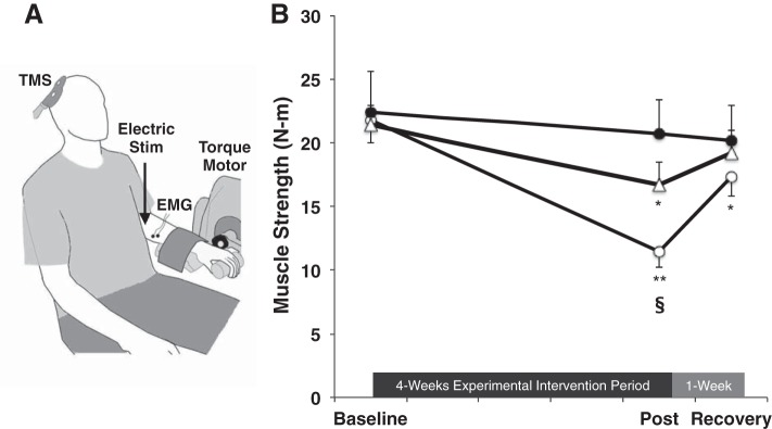
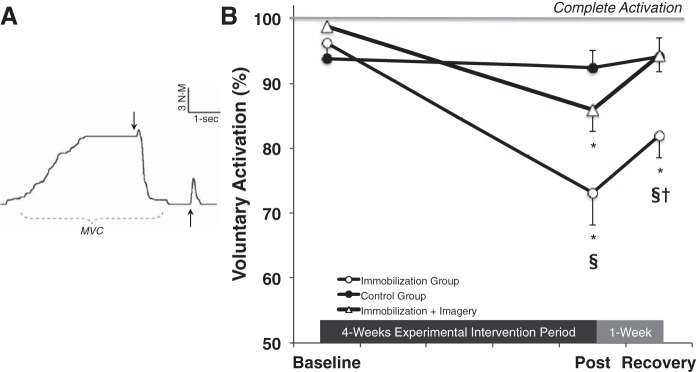
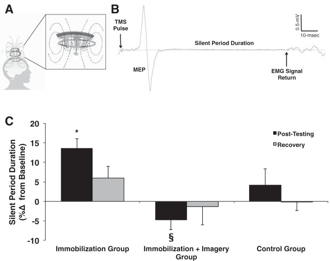
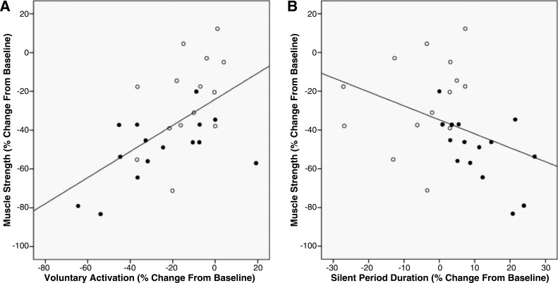
We tested the hypothesis that the nervous system, and the cortex in particular, is a critical determinant of muscle strength/weakness and that a high level of corticospinal inhibition is an important neurophysiological factor regulating force generation. A group of healthy individuals underwent 4 wk of wrist-hand immobilization to induce weakness. Another group also underwent 4 wk of immobilization, but they also performed mental imagery of strong muscle contractions 5 days/wk. Mental imagery has been shown to activate several cortical areas that are involved with actual motor behaviors, including premotor and M1 regions. A control group, who underwent no interventions, also participated in this study. Before, immediately after, and 1 wk following immobilization, we measured wrist flexor strength, voluntary activation (VA), and the cortical silent period (SP; a measure that reflect corticospinal inhibition quantified via transcranial magnetic stimulation). Immobilization decreased strength 45.1 ± 5.0%, impaired VA 23.2 ± 5.8%, and prolonged the SP 13.5 ± 2.6%. Mental imagery training, however, attenuated the loss of strength and VA by ∼50% (23.8 ± 5.6% and 12.9 ± 3.2% reductions, respectively) and eliminated prolongation of the SP (4.8 ± 2.8% reduction). Significant associations were observed between the changes in muscle strength and VA (r = 0.56) and SP (r = -0.39). These findings suggest neurological mechanisms, most likely at the cortical level, contribute significantly to disuse-induced weakness, and that regular activation of the cortical regions via imagery attenuates weakness and VA by maintaining normal levels of inhibition.

Keywords: dynapenia; imagery; immobilization; muscle; strength; weakness.


Figures

Fig. 1.
Fig. 1.
A: setup for assessing wrist flexion strength, voluntary activation (VA), and the cortical silent period (SP). TMS, transcranial magnetic stimulation; EMG, electromyogram. B: immobilization (open circles; n = 15) resulted in a 45% reduction in strength. Mental imagery training (open triangles; n = 14), however, attenuated the loss of muscle strength by ∼50% (strength loss of 24%). No changes were observed in the control group (solid circles; n = 15). Values are means ± SE. Significant differences vs. *baseline, **baseline and recovery, §control group value.
Fig. 2.
Fig. 2.
A: example of a force trace assessing VA. Arrows represent the delivery of a 100-Hz electrical doublet to the peripheral nerve while an individual is maximally contracting (first arrow) and ∼2 s after the completion of the contraction (second arrow). MVC, maximum voluntary contraction. B: immobilization (open circles; n = 15) reduced VA ∼25%. Mental imagery training (open triangles; n = 14), however, attenuated the impairment in VA by ∼50%. No changes were observed in the control group (solid circles; n = 15). Values are means ± SE. Significant differences vs. *baseline, §control group value, †imagery group value.
Fig. 3.
Fig. 3.
A: the TMS coil induces a magnetic field and a subsequent Eddy current that stimulates neurons within the motor cortex. B: example of an EMG trace illustrating a motor evoked potential (MEP) and SP. In this study, single TMS pulses were delivered to the primary motor cortex during 15% of maximum contraction to quantify the SP duration as an index of GABAB-mediated inhibition. C: immobilization resulted in a 12% prolongation in the SP (n = 15). Mental imagery training (n = 14), however, eliminated prolongation of the SP. No changes were observed in the control group (n = 15). Values are presented as a %change for clarity, but it should be noted that no baseline differences in groups were observed (baseline measures for control group: 108.5 ± 4.3 ms, immobilization group was 107.5 ± 4.4 ms, and immobilization + imagery group was 110.5 ± 4.9 ms). Values are means ± SE. Significant differences vs. *baseline, §immobilization group value.
Fig. 4.
Fig. 4.
A: there was a positive association between the percent change in muscle strength and the percent change in VA following 4 wk of cast immobilization (solid symbols: immobilization group; open symbols: immobilization + imagery group). This finding indicates that individuals who experienced the largest immobilization-induced loss of muscle strength also experienced the largest immobilization-induced impairments in voluntary (neural) activation (r = 0.56, P < 0.01). B: there was a negative association between the percent change in muscle strength and the percent change in the cortical SP duration following 4 wk of cast immobilization (solid symbols: immobilization group; open symbols: immobilization + imagery group). This finding indicates that individuals who experienced the largest immobilization-induced loss of muscle strength also experienced the largest immobilization-induced prolongation in the cortical SP (r = −0.39, P = 0.03).

References

    1. Adkins DL, Boychuk J, Remple MS, Kleim JA. Motor training induces experience-specific patterns of plasticity across motor cortex and spinal cord. J Appl Physiol 101: 1776–1782, 2006. - PubMed
    1. Aoyama T, Kaneko F. The effect of motor imagery on gain modulation of the spinal reflex. Brain Res 1372: 41–48, 2011. - PubMed
    1. Ashe J. Force and the motor cortex. Behav Brain Res 87: 255–269, 1997. - PubMed
    1. Bakker M, Overeem S, Snijders AH, Borm G, van Elswijk G, Toni I, Bloem BR. Motor imagery of foot dorsiflexion and gait: effects on corticospinal excitability. Clin Neurophysiol 119: 2519–2527, 2008. - PubMed
    1. Butler JE, Petersen NC, Herbert RD, Gandevia SC, Taylor JL. Origin of the low-level EMG during the silent period following transcranial magnetic stimulation. Clin Neurophysiol 123: 1409–1414, 2012. - PubMed

Publication types

LinkOut - more resources