. 2014 Oct 1;28(19):2120-33.
doi: 10.1101/gad.244749.114.

Network dynamics determine the autocrine and paracrine signaling functions of TNF

Affiliations

Network dynamics determine the autocrine and paracrine signaling functions of TNF

Andrew B Caldwell et al. Genes Dev. .

Abstract

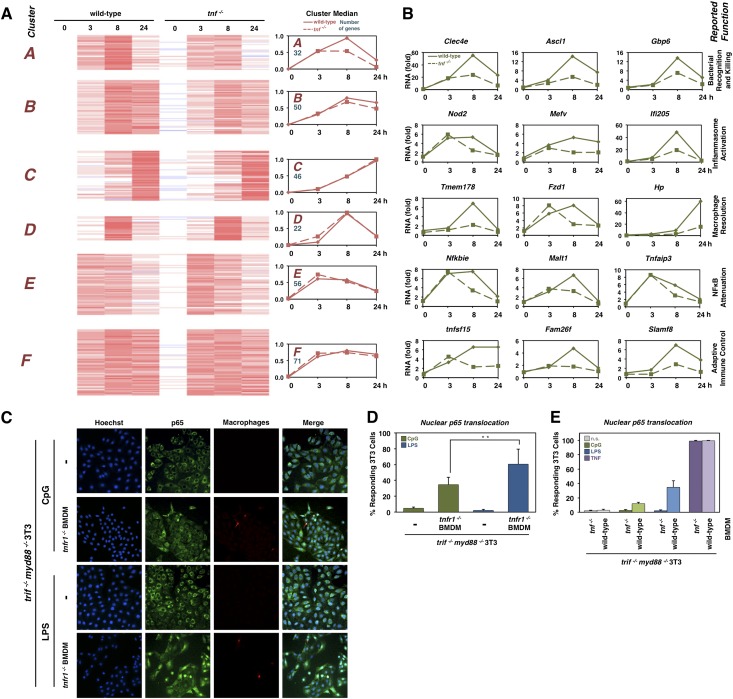
A hallmark of the inflammatory response to pathogen exposure is the production of tumor necrosis factor (TNF) that coordinates innate and adaptive immune responses by functioning in an autocrine or paracrine manner. Numerous molecular mechanisms contributing to TNF production have been identified, but how they function together in macrophages remains unclear. Here, we pursued an iterative systems biology approach to develop a quantitative understanding of the regulatory modules that control TNF mRNA synthesis and processing, mRNA half-life and translation, and protein processing and secretion. By linking the resulting model of TNF production to models of the TLR-, the TNFR-, and the NFκB signaling modules, we were able to study TNF's functions during the inflammatory response to diverse TLR agonists. Contrary to expectation, we predicted and then experimentally confirmed that in response to lipopolysaccaride, TNF does not have an autocrine function in amplifying the NFκB response, although it plays a potent paracrine role in neighboring cells. However, in response to CpG DNA, autocrine TNF extends the duration of NFκB activity and shapes CpG-induced gene expression programs. Our systems biology approach revealed that network dynamics of MyD88 and TRIF signaling and of cytokine production and response govern the stimulus-specific autocrine and paracrine functions of TNF.

Keywords: MAP kinase; NFκB; inflammation; innate immune response; pathogen sensors.


Figures

Figure 1.
Figure 1.
TNF production is regulated by both MyD88 and TRIF pathways, but only NFκB, not IRF3/7, is required for its transcriptional induction. (A) Diagram illustrating molecular mechanisms potentially regulating the production of TNF; solid lines indicate mechanisms known to play a role in macrophages, and dashed lines indicate mechanism that have been reported in the literature in different cell systems. (B) Schematic of the three regulatory modules that control TNF expression, highlighting the experimentally quantifiable input and output of each module. (C) Secretion of TNF as measured by ELISA of cell media from wild-type, trif −/−, or myd88−/− bone marrow-derived macrophages (BMDMs). Cells were stimulated with 10 ng/mL LPS; n = 3. (D) Levels of TNF mRNA (log2 fold) produced by wild-type, trif−/−, or myd88−/− BMDMs stimulated with 10 ng/mL LPS, measured by RT-qPCR. Wild type, n = 5. trif−/−, n = 3. myd88−/−, n = 3. (E) Levels of nascent TNF RNA (log2 fold) produced by wild-type, trif−/−, or myd88−/− BMDMs stimulated with 10 ng/mL LPS, measured by RT–PCR. Wild type, n = 3. Nascent transcripts measured by RT-qPCR with intron–exon junction-spanning primers. (F) TNF mRNA levels (fold) measured by RT-qPCR in wild-type or rela−/−relb−/−crel−/− fetal liver-derived macrophages (FLDMs) stimulated with 100 ng/mL LPS. (G) Levels of TNF mRNA (log2 fold) produced by wild-type or irf3−/−irf7−/− BMDMs stimulated with 10 ng/mL LPS, measured by RT–PCR (wild type, n = 5; irf3−/−irf7-/-, n = 3). (H) Levels of nascent TNF RNA (log2 fold) produced by wild-type or irf3−/−irf7−/− BMDMs stimulated with 10 ng/mL LPS, measured by RT-qPCR (n = 3). (I) Quantified activation of NFκB measured by EMSA (κB-site-containing HIV probe) in wild-type, trif−/−, or myd88−/− BMDMs stimulated with 10 ng/mL LPS. Quantification of NFκB EMSA bands normalized to peak activity (n = 3). (J) Schematic of regulatory module 1 that determines nascent TNF mRNA; input is quantified NFκB activation data (shown in H), and output is nascent TNF mRNA. (K) Levels of nascent mRNA simulated by the mathematical model in I (solid lines) and determined experimentally in E (data points) for wild-type, trif−/−, or myd88−/− genotypes. For all graphs, error bars indicate one standard deviation. The combined root mean square difference (RMSD) between all model simulations and experimental data points shown are indicated.
Figure 2.
Figure 2.
TRIF regulates TNF mRNA half-life and translation and protein processing and secretion. (A) TNF mRNA half-life measured by RT-qPCR following actinomycin-D time course in wild-type, trif−/−, or myd88−/− BMDMs prestimulated for 30 min with 10 ng/mL TNF alone (purple) or 10 ng/mL TNF and 10 ng/mL LPS (blue) (wild-type, n = 5; trif−/−, n = 4; myd88−/−, n = 3). (B) TNF mRNA half-life measured as in A in wild-type BMDMs following prestimulation with 10 ng/mL TNF alone (purple); 10 ng/mL TNF and 10 ng/mL LPS (blue); or 10 ng/mL TNF, 10 ng/mL LPS, and 10µM p38 inhibitor for 30 min (white) (TNF, n = 5; LPS, n = 5; p38, n = 1). (C) Immunoblots for phospho-p38, phospho-ERK, phospho-MK2, and actin of whole-cell extracts made from wild-type, trif−/−, or myd88−/− BMDMs stimulated with 10 ng/mL LPS. Blots shown are representative of four experiments. (Below) Quantification of immunoblots normalized to wild-type peak phosphorylation. Error bars indicate one standard deviation; (*) P-value < 0.05; (**) P-value < 0.02 for difference between wild-type and trif−/− time points. (D) Immunoblots for phospho-MK2, phospho-TTP, and actin of whole-cell extracts made from wild-type BMDMs pretreated with DMSO, 10 µM p38 inhibitor, or 10 µM ERK inhibitor for 1 h followed by stimulation with 10 ng/mL LPS. (E) Schematic of regulatory module 2 that controls TNF mRNA half-life via TRIF. (Input) Nascent TNF mRNA experimental data; (output) mature TNF mRNA abundance. (F) Computational simulations of module 2 (solid lines) for TNF mRNA production in the wild-type, trif−/−, or myd88−/− genotype in response to 10 ng/mL LPS with either no stabilization control (top left), stabilization by TRIF and MyD88 (top right), stabilization by MyD88 alone (bottom left), or stabilization by TRIF alone (bottom right). Data points indicate experimental data for TNF mRNA in wild-type, trif−/−, or myd88−/− BMDMs stimulated with 10 ng/mL LPS as reported in Figure 1C. The combined RMSD between model simulations for each half-life control mechanism and experimental data points shown are indicated.
Figure 3.
Figure 3.
TRIF regulates TNF translation, protein processing, and secretion. (A) Immunoblot for pro-TNF and actin in wild-type, trif−/−, and myd88−/− BMDMs stimulated with 10 ng/mL LPS. Data are representative of three experiments. (Right) Quantification of pro-TNF bands normalized to peak wild-type protein levels. Error bars indicate one standard deviation; (*) P-value < 0.05; (**) P-value < 0.02 for difference between wild-type and trif−/− time points. (B) Immunoblot for pro-TNF and actin of whole-cell extracts from wild-type BMDMs pretreated with DMSO, 10 µM p38 inhibitor, or 10 µM ERK inhibitor for 1 h followed by stimulation with 10 ng/mL LPS. The blot is representative of two experiments. (C) Immunoblot for phospho-eIF4E, phospho-TACE, and actin of whole-cell extracts from wild-type, trif−/−, and myd88−/− BMDMs stimulated with 10 ng/mL. Phospho-eIF4E, n = 2; p-TACE, n = 3. (Below) Quantification of p-eIF4E and p-TACE immunoblot bands normalized to peak wild-type levels; (*) P-value < 0.05 for a difference between wild-type and trif−/− time points. (D) Immunoblot for phospho-eIF4E, eIF4E, phospho-TACE, TACE, and actin of whole-cell extracts from wild-type BMDMs pretreated with DMSO, 10 µM p38 inhibitor, or 10 µM ERK inhibitor for 1 h followed by stimulation with 10 ng/mL LPS. The blots are representative of two experiments. (E) Schematic of regulatory module 3 describing the promotion of TNF translation and secretion by TRIF. (Input) Experimental data for mature TNF mRNA; (output) secreted TNF. (F) Computational simulations (solid lines) of module 3 for pro-TNF expression without (left) and with (right) the promotion of TNF procession through TRIF-mediated translation regulation. Data points indicate experimental data for pro-TNF expression in wild-type, trif−/−, or myd88−/− BMDMs stimulated with 10 ng/mL LPS as reported in A, respectively. RMSD between model simulations and experimental data points are indicated for each genotype. (G) Computational simulations (solid lines) of module 3 for secreted TNF without (left) and with (right) the promotion of TNF secretion through TRIF-mediated secretion regulation. Data points indicate experimental data for TNF secretion in wild-type, trif−/−, or myd88−/− BMDMs stimulated with 10 ng/mL LPS as reported in Figure 1C, respectively.
Figure 4.
Figure 4.
A multimodular model of the TNF production network accounts for experimental data for some TLR agonists but not others. (A) Schematic of the computational model combining models of the regulatory modules for TLR activation of adaptors TRIF and MyD88 converging on IKK, activation of NFκB by IKK, and the three modules for TNF production. (B) Model simulations and experimental data for nascent mRNA, mature mRNA, pro-TNF, and secreted TNF protein in wild-type cells in response to 10 ng/mL LPS; solid lines indicate values of model simulations, and points represent experimental data represented in previous figures. (C) Model simulations and experimental data for mature TNF mRNA and secreted TNF for wild-type cells in response to 500 nM CpG and 50 µg/mL PolyI:C. (D) Model simulations and experimental data for same molecular species for trif−/− and myd88−/− cells in response to 10 ng/mL LPS. For experimental data points, n = 3. Error bars are one standard deviation from the mean. RMSDs are indicated for each panel in B–D.
Figure 5.
Figure 5.
TLR-responsive TNF production functions in an autocrine manner in response to some TLR agonists but not others. (A) Schematic of the expanded multimodular computational model, incorporating the TNFR-to-NFκB signaling module. (B) Model simulations and experimental data for mature TNF mRNA and secreted TNF protein in wild-type cells stimulated with 10 ng/mL LPS, 500 nM CpG, or 50 µg/mL PolyI:C; solid lines indicate values of model simulations, and points represent experimental data presented in previous figures. RMSDs are shown for each panel. (C) Model simulations for NFκB activity in wild-type or tnf−/− stimulated by 10 ng/mL LPS or 100 nM CpG. Solid lines indicate wild-type simulation, and dashed lines indicate tnf−/−. (D) Experimental validation of model simulations in C. Activation of NFκB measured by EMSA in wild-type and tnf−/− BMDMs stimulated with 10 ng/mL LPS or 100 nM CpG. Graphs are quantification of experimental data normalized to peak wild-type NFκB activation (n = 4). The gel image is representative of four experiments. (**) P-value < 0.02 for a difference between wild-type and tnf−/− time points.
Figure 6.
Figure 6.
TNF’s paracrine- and autocrine-specific functions are TLR-specific. (A) Transcriptomic analysis of wild-type and tnf−/− BMDMs responding to 100 nM CpG. K-means clustering of 267 gene expression profiles. (Right) Cluster median reveals TNF dependency of clusters A and B. (B) Select genes whose expression is TNF-dependent in response to CpG, grouped by their known roles in inflammation and macrophage function. (C) Immunofluorescence images of coculture of tnfr1−/− BMDMs with trif−/−myd88−/− 3T3s. The top row shows p65 staining of trif−/−myd88−/− 3T3s stimulated with 1 µM CpG for 75 min. The second row shows p65 staining of coculture of tnfr−/− BMDMs with trif−/−myd88−/− 3T3s stimulated with 1 µM CpG for 75 min. The third row shows p65 staining of trif−/−myd88−/− 3T3s stimulated with 1 µg/mL LPS for 75 min. The fourth row shows p65 staining of coculture of tnfr−/− BMDMs with trif−/−myd88−/− 3T3s stimulated with 1 µg/mL LPS for 75. Images are representative of three separate experiments. (D) Bar graphs showing the average percentage of 3T3 cells with nuclear p65 in conditions described in C. Three-hundred to 500 cells were counted over three experiments. Error bars indicate standard error of the mean. (**) P-value = 0.02. (E) Bar graphs of the percentage of trif−/−myd88−/− 3T3 cells with nuclear p65 when cocultured with wild-type or tnf−/− BMDMs and stimulated as indicated. More than 1000 cells were counted over three experiments.

References

    1. Alexopoulou L, Holt AC, Medzhitov R, Flavell RA. 2001. Recognition of double-stranded RNA and activation of NF-κB by Toll-like receptor 3. Nature 413: 732–738 - PubMed
    1. Alexopoulou L, Kranidioti K, Xanthoulea S, Denis M, Kotanidou A, Douni E, Blackshear PJ, Kontoyiannis DL, Kollias G. 2006. Transmembrane TNF protects mutant mice against intracellular bacterial infections, chronic inflammation and autoimmunity. Eur J Immunol 36: 2768–2780 - PubMed
    1. Anders S, Pyl PT, Huber W. 2014. HTSeq—a Python framework to work with high-throughput sequencing data. bioRxiv doi: 10.1101/002824 - DOI - PMC - PubMed
    1. Andersson K, Sundler R. 2006. Posttranscriptional regulation of TNFα expression via eukaryotic initiation factor 4E (eIF4E) phosphorylation in mouse macrophages. Cytokine 33: 52–57 - PubMed
    1. Basak S, Behar M, Hoffmann A. 2012. Lessons from mathematically modeling the NF-κB pathway. Immunol Rev 246: 221–238 - PMC - PubMed

Publication types

MeSH terms

Substances