. 2014 Oct 1;28(19):2175-87.
doi: 10.1101/gad.216226.113.

The Prox1-Vegfr3 feedback loop maintains the identity and the number of lymphatic endothelial cell progenitors

Affiliations

The Prox1-Vegfr3 feedback loop maintains the identity and the number of lymphatic endothelial cell progenitors

R Sathish Srinivasan et al. Genes Dev. .

Abstract

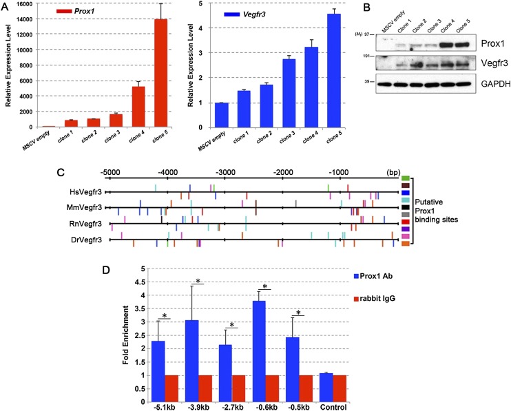
The mammalian lymphatic vasculature is important for returning fluids from the extracellular tissue milieu back to the blood circulation. We showed previously that Prox1 dosage is important for the development of the mammalian lymphatic vasculature. The lack of Prox1 activity results in the complete absence of lymphatic endothelial cells (LECs). In Prox1 heterozygous embryos, the number of LECs is reduced because of a decrease in the progenitor pool in the cardinal vein. This reduction is caused by some progenitor cells being unable to maintain Prox1 expression. In this study, we identified Vegfr3, the cognate receptor of the lymphangiogenic growth factor Vegfc, as a dosage-dependent, direct in vivo target of Prox1. Using various mouse models, we also determined that Vegfr3 regulates Prox1 by establishing a feedback loop necessary to maintain the identity of LEC progenitors and that Vegfc-mediated activation of Vegfr3 signaling is necessary to maintain Prox1 expression in LEC progenitors. We propose that this feedback loop is the main sensing mechanism controlling the number of LEC progenitors and, as a consequence, the number of budding LECs that will form the embryonic lymphatic vasculature.

Keywords: Prox1; Vegfr3; endothelial cell; lymphatics; mouse; progenitor.


Figures

Figure 1.
Figure 1.
Vegfr3 is a dosage-dependent target of Prox1. (A) H5V cells were infected with Prox1-expressing retroviruses. Individual clones were picked following positive selection with antibiotics. RNA extracted from the clones was analyzed by real-time PCR for the expression of Prox1 (left graph) and Vegfr3 (right graph). (B) Protein levels of Vegfr3 appear to correlate directly with those of Prox1. (C) DNA regions located 5 kb upstream of exon 1 of human (Hs), mouse (Mm), rat (Rn), and zebrafish (Dr) Vegfr3 were analyzed for putative Prox1-binding sites by TRANSFAC bioinformatics software. Upward bars indicate the sites in the sense orientation, and downward bars indicate those in the antisense orientation. Colored boxes identify the different Prox1 recognition sites (see Supplemental Table 1). (D) ChIP was carried out on mouse LECs collected by flow cytometry. A rabbit polyclonal antibody against Prox1 or control IgG was used to pull down the DNA fragments. Real-time PCR was carried out using primers specific for the various regions along the 5-kb DNA element of mouse Vegfr3. Substantial Prox1 binding was observed at the indicated sites. These data are from three independent experiments. (*) P < 0.05.
Figure 2.
Figure 2.
LEC progenitors and LECs are reduced in Prox1+/GFPCre;Vegfr3+/LacZ embryos. Vegfr3+/LacZ mice were bred with Prox1+/GFPCre to generate Prox1+/GFPCre;Vegfr3+/LacZ double-heterozygous embryos. (A–C) At E13.5 Vegfr3+/LacZ embryos were phenotypically normal (A), Prox1+/GFPCre embryos had mild edema (B), and severe edema and regions of hemorrhagic accumulation were observed in most Prox1+/GFPCre;Vegfr3+/LacZ embryos (C). Prox1+/GFPCre and Prox1+/GFPCre;Vegfr3+/LacZ embryos were collected at E11.5 (D–F), E13.5 (G,H), or E15.5 (I,J) and analyzed by immunohistochemistry for the LEC markers Prox1 and podoplanin together with the pan-EC marker PECAM1. (D,E,G,H) Transverse sections, with the neural tube and heart at the right and left, respectively, of the figures. (I,J) Whole mounts on the peripheral skin. (D,E) Compared with Prox1+/GFPCre embryos, the expression of Prox1 and podoplanin is reduced in Prox1+/GFPCre;Vegfr3+/LacZ littermates at E11.5. (F) Furthermore, the number of Prox1+ LECs is reduced in Prox1+/GFPCre;Vegfr3+/LacZ embryos at E11.5 (n = 3 for each genotype). At E13.5, while a clear lymph sac (LS) was seen in Prox1+/GFPCre embryos (G), scattered and mispatterned blood-filled structures were seen in Prox1+/GFPCre;Vegfr3+/LacZ embryos (H; arrows). At E15.5, lymphatic vessels are seen in Prox1+/GFPCre embryonic skin (I); however, in Prox1+/GFPCre;Vegfr3+/LacZ embryos, the few Prox1+ structures (dashed lines) failed to express podoplanin (J). P-values are as follows: (**) P = 0.0068; (*) P = 0.0128. Bars, 100 μm.
Figure 3.
Figure 3.
Prox1+/GFPCre;Vegfr3+/LacZ embryos fail to maintain Prox1 expression in LEC progenitors. Prox1+/GFPCre mice were bred with Vegfr3+/LacZ;R26mT/mG mice, and the resulting E10.5 Prox1+/GFPCre;R26mT/mG and Prox1+/GFPCre; Vegfr3+/LacZ;R26mT/mG embryos were analyzed by immunohistochemistry for Prox1 and GFP. Upon Cre-mediated activation, the R26mT/mG reporter expresses a membrane-tagged GFP that allows the visualization of the cell surface. (AC) In Prox1+/GFPCre;R26mT/mG embryos, numerous Prox1+ cells were seen on the CV. All of these cells and a few Prox1 cells on the CV were GFP+ (white dotted line). (D–F) In contrast, in Prox1+/GFPCre; Vegfr3+/LacZ;R26mT/mG embryos, fewer Prox1+ cells were seen on the CV, although most of these cells were GFP+ (white dotted line). Bars, 100 μm. (G) For cell counting, the number of GFP+ Prox1DAPI+ or GFP+ Prox1+DAPI+ cells were quantitated in stained sections (n = 3 embryos for each genotype). Cell counting showed that the number of Prox1 GFP+ LEC progenitors (P-value = 0.065) was higher in double heterozygous, indicating that the Vegfr3–Prox1 interaction is required to maintain LEC fate.
Figure 4.
Figure 4.
Vegfr3 regulates Prox1 expression in LEC progenitors and differentiating LECs. Wild-type (WT) [Tg(Prox1-tdTomato)], Prox1+/−[Tg(Prox1-tdTomato);Prox1+/LacZ], and Vegfr3+/− [Tg(Prox1-tdTomato); Vegfr3+/LacZ] embryos were collected at E11.5 (one embryo per genotype). Using laser capture microdissection, LEC progenitors (inside the CV) and differentiating LECs (outside the CV) were collected separately, and the levels of Prox1 (A) and Vegfr3 (B) were analyzed by quantitative real-time PCR. mRNA levels are shown as fold of increase and were normalized to PECAM1 levels. These qPCR results were generated from a single laser capture experiment using pooled mRNA extracted from cells captured from 10-μm sections obtained from the entire embryo for each genotype.
Figure 5.
Figure 5.
Prox1 and Vegfr3 levels are reduced in Prox1+/GFPCre;Vegfr3+/LacZ embryos. (A–H′) Immunohistochemistry for Prox1, Vegfr3, and PECAM1 was carried out in E11.5 wild-type (WT) (A,A′,E,E′), Vegfr3+/LacZ (B,B′,F,F′), Prox1+/GFPCre (C,C′,G,G′), and Prox1+/GFPCre;Vegfr3+/LacZ (D,D′,H,H′) embryos. The expression levels of Prox1, Vegfr3, and PECAM1 in venous LEC progenitors (A–D′) and differentiating LECs outside of the CV (E–H′) were quantified using the Slidebook image analysis software (I,J). Prox1 and Vegfr3 expression levels were significantly reduced in Prox1+/GFPCre;Vegfr3+/LacZ embryos compared with their single heterozygous littermates. At least three embryos for each genotype were used for quantification. Bars: A–H, 100 μm.
Figure 6.
Figure 6.
Feedback loop regulation between Prox1 and Vegfr3 is at the RNA and protein levels. (A) hLECs were transfected with siRNAs and cultured in conditioned medium. After 48 hm mRNA was extracted, and qPCR was carried out to quantify Prox1 and Vegfr3 expression levels. siProx1 down-regulates both Prox1 and Vegfr3 levels (P < 0.0001 and P = 0.0108, respectively). Likewise, siVegfr3 down-regulates both Vegfr3 and Prox1 levels (P = 0.0003 and P = 0.0051, respectively). These data are the result of three independent experiments. (B) hLECs transfected with siRNA were cultured and immunostained for Prox1. Prox1 expression was down-regulated in those cells that were successfully transfected with fluorescently labeled siRNAs directed against either Prox1 or Vegfr3 (arrowheads).
Figure 7.
Figure 7.
Prox1 and Vegfc genetically interact with each other to regulate the number of LEC progenitors and LECs. (A–D) Prox1+/GFPCre and Vegfc+/− mice were bred with each other, and embryos were collected at E11.5 and analyzed by immunohistochemistry for the LEC markers Prox1 and podoplanin together with the pan-EC marker PECAM1. Compared with wild-type (WT) embryos (A), Vegfc+/− (B) and Prox1+/GFPCre (C) embryos had fewer Prox1+ LEC progenitors and LECs. (D) Prox1+/GFPCre;Vegfc+/− embryos had even fewer LECs. (E) Quantification of the cell numbers revealed that the differences in differentiating LEC numbers are statistically significant ([***] P ≤ 0.001; n = 3 embryos for each genotype). Regarding LEC progenitors, there is no significant difference; however, there is a trend that shows that the number of LECs decreases in the double-heterozygous embryos (P = 0.0925). Bar, 100 μm.
Figure 8.
Figure 8.
Model of the feedback loop regulating the specification of Prox1-expressing LEC progenitors in the embryonic veins. Between E9.75 and E13.5, in some venous EC cells with low or no Notch signaling, CoupTFII is up-regulated; then, in combination with Sox18, it induces Prox1 expression such that LEC progenitors start to be specified. Notch signaling is repressed in the specified LEC progenitors by Coup-TFII. Prox1 in turn activates the expression of Vegfr3 in a dosage-dependent manner. Activation of Vegfr3 signaling by Vegfc will also maintain Prox1 expression in LEC progenitors and differentiating LECs. Coup-TFII also interacts with Prox1 to maintain Prox1 expression. Coup-TFII and Prox1 also likely maintain Notch signaling at low levels in LEC progenitors at this stage. Prox1+ LEC progenitors will subsequently bud off from the CV and intersomitic vessels (ISV) and start to express differentiation markers such as podoplanin. After E13.5, Notch signaling levels are increased in venous ECs such that Notch will suppress Coup-TFII to prevent further specification of Prox1+ LEC progenitors. At the same time, Notch signaling also likely inhibits Vegfr3 expression via Hey1/2, thereby short-circuiting the Prox1–Vegfr3 feedback loop and stopping the generation of LEC progenitors in the veins. It could be speculated that most likely the Prox1–Vegfr3 feedback loop does not operate in differentiated LECs.

References

    1. Aranguren XL, Beerens M, Coppiello G, Wiese C, Vandersmissen I, Nigro AL, Verfaillie CM, Gessler M, Luttun A. 2013. COUP-TFII orchestrates venous and lymphatic endothelial identity by homo- or heterodimerisation with PROX1. J Cell Sci 126: 1164–1175 - PubMed
    1. Chen CY, Bertozzi C, Zou Z, Yuan L, Lee JS, Lu M, Stachelek SJ, Srinivasan S, Guo L, Vicente A, et al. 2012. Blood flow reprograms lymphatic vessels to blood vessels. J Clin Invest 122: 2006–2017 - PMC - PubMed
    1. Dumont DJ, Jussila L, Taipale J, Lymboussaki A, Mustonen T, Pajusola K, Breitman M, Alitalo K. 1998. Cardiovascular failure in mouse embryos deficient in VEGF receptor-3. Science 282: 946–949 - PubMed
    1. Flister MJ, Wilber A, Hall KL, Iwata C, Miyazono K, Nisato RE, Pepper MS, Zawieja DC, Ran S. 2010. Inflammation induces lymphangiogenesis through up-regulation of VEGFR-3 mediated by NF-κB and Prox1. Blood 115: 418–429 - PMC - PubMed
    1. Francois M, Caprini A, Hosking B, Orsenigo F, Wilhelm D, Browne C, Paavonen K, Karnezis T, Shayan R, Downes M, et al. 2008. Sox18 induces development of the lymphatic vasculature in mice. Nature 456: 643–647 - PubMed

Publication types

MeSH terms

Substances

LinkOut - more resources