. 2014 Oct 29;42(19):11921-7.
doi: 10.1093/nar/gku896. Epub 2014 Oct 1.

A biomechanical mechanism for initiating DNA packaging

Affiliations

A biomechanical mechanism for initiating DNA packaging

Haowei Wang et al. Nucleic Acids Res. .

Abstract

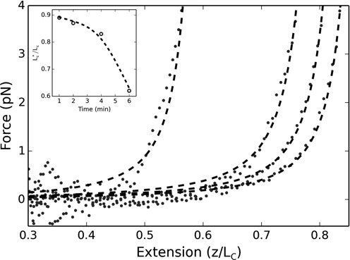
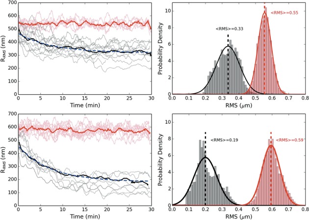
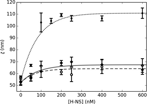
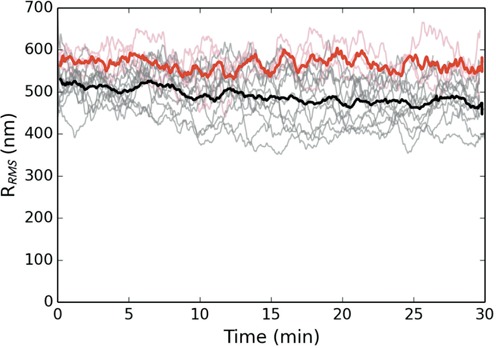
The bacterial chromosome is under varying levels of mechanical stress due to a high degree of crowding and dynamic protein-DNA interactions experienced within the nucleoid. DNA tension is difficult to measure in cells and its functional significance remains unclear although in vitro experiments have implicated a range of biomechanical phenomena. Using single-molecule tools, we have uncovered a novel protein-DNA interaction that responds to fluctuations in mechanical tension by condensing DNA. We combined tethered particle motion (TPM) and optical tweezers experiments to probe the effects of tension on DNA in the presence of the Hha/H-NS complex. The nucleoid structuring protein H-NS is a key regulator of DNA condensation and gene expression in enterobacteria and its activity in vivo is affected by the accessory factor Hha. We find that tension, induced by optical tweezers, causes the rapid compaction of DNA in the presence of the Hha/H-NS complex, but not in the presence of H-NS alone. Our results imply that H-NS requires Hha to condense bacterial DNA and that this condensation could be triggered by the level of mechanical tension experienced along different regions of the chromosome.


Figures

Figure 1.
Figure 1.
Force-extension curves for 200 nM of H-NS and Hha. From right to left: 1, 2, 4 and 6 min after optically trapping/centering the attached microsphere. Dashed curves are WLC fits with variable DNA persistence and contour lengths. Inset: relative change in contour length obtained by the fits (dashed line is an interpolation).
Figure 2.
Figure 2.
Dynamics of DNA condensation after applied tension. The top (bottom) figure is for equal concentrations of H-NS and Hha of 100 (200) nM. The red curves are the control absent an force applied. The prominent solid lines are averaged over the respective trajectories and fitted by a decaying exponential (dashed/blue) (τ = 7.5 (7.3) min for 100 (200) nM protein). All trajectories have been smoothed by a 60 s running window. Top (bottom) right: RMS histograms for 100 (200) nM protein for control (red) and collapsed (black) population after 15 minutes. The trajectories were fitted to a Gaussian to provide a mean RMS value.
Figure 3.
Figure 3.
Persistence length dependence upon H-NS concentration. Measured via TPM pre-stressed (circles/dotted line) and post stressed (open circles/dashed line). Measured via force extension with optical tweezers (diamonds/solid line). Exponential fits to the data (lines) provide a reasonable interpolation.
Figure 4.
Figure 4.
Dynamics of DNA bound by H-NS and the mutant Hha(R14A/R17A). The black curves display the RMS excursion of the tethered microspheres as a function of time after release from the optical trap. The red curves are a control absent an applied force. Both proteins are at a concentration of 200 nM. The prominent solid curves are averaged over the respective trajectories for each population. All trajectories have been smoothed by a 60 s running window. While the mutant protein tends to reduce the RMS observed, the protein–DNA complex can no longer be triggered to collapse.
Figure 5.
Figure 5.
Dynamics of DNA bound by 200 nM of H-NS and 2 mM of MgCl2 as a function of time. The black curves display the RMS excursion of the tethered microspheres as a function of time after release from the optical trap. The red curves are a control absent an applied force. The prominent solid curves are averaged over the respective trajectories for each population. All trajectories have been smoothed by a 60 s running window. Notice that the DNA, bound by H-NS alone, can be triggered to collapse in the presence of millimolar concentrations of Mg2+.
Figure 6.
Figure 6.
(A) Schematic of an H-NS oligomer in complex with Hha (the positively charged surface of Hha is indicated by ‘+’). (B) By dissociating one protein at a time, this bridging configuration should be relatively sensitive to an applied force (indicated by the arrows). (C) An alternative orientation that would be much more stable to dissociation by an applied tension.

References

    1. Ochman H., Lawrence J.G., Groisman E.A. Lateral gene transfer and the nature of bacterial innovation. Nature. 2000;405:299–304. - PubMed
    1. Gevers D., Cohan F.M., Lawrence J.G., Spratt B.G., Coenye T., Feil E.J., Stackebrandt E., Van de Peer Y., Vandamme P., Thompson F.L., et al. Opinion: re-evaluating prokaryotic species. Nat. Rev. Microbiol. 2005;3:733–739. - PubMed
    1. Jain R., Rivera M.C., Moore J.E., Lake J.A. Horizontal gene transfer in microbial genome evolution. Theor. Popul. Biol. 2002;61:489–495. - PubMed
    1. Jun S., Wright A. Entropy as the driver of chromosome segregatioin. Nat. Rev. Microbiol. 2010;8:600–607. - PMC - PubMed
    1. Thanbichler M., Wang S.C., Shapiro L. The bacterial nucleoid: a highly organized and dynamic structure. J. Cell. Biochem. 2005;96:506–521. - PubMed

Publication types