Review
. 2014 Oct 1;4(10):a015172.
doi: 10.1101/cshperspect.a015172.

The genetics of human skin disease

Affiliations
Review

The genetics of human skin disease

Gina M DeStefano et al. Cold Spring Harb Perspect Med. .

Abstract

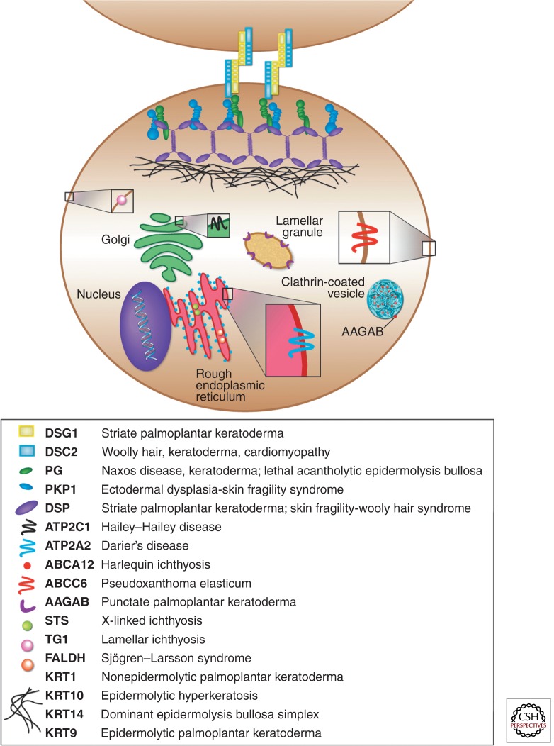
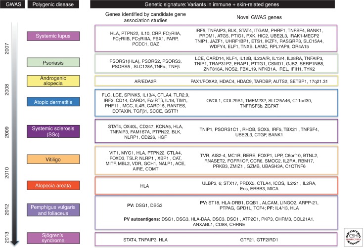
The skin is composed of a variety of cell types expressing specific molecules and possessing different properties that facilitate the complex interactions and intercellular communication essential for maintaining the structural integrity of the skin. Importantly, a single mutation in one of these molecules can disrupt the entire organization and function of these essential networks, leading to cell separation, blistering, and other striking phenotypes observed in inherited skin diseases. Over the past several decades, the genetic basis of many monogenic skin diseases has been elucidated using classical genetic techniques. Importantly, the findings from these studies has shed light onto the many classes of molecules and essential genetic as well as molecular interactions that lend the skin its rigid, yet flexible properties. With the advent of the human genome project, next-generation sequencing techniques, as well as several other recently developed methods, tremendous progress has been made in dissecting the genetic architecture of complex, non-Mendelian skin diseases.


Figures

Figure 1.
Figure 1.
The classical genetic approach of functional and positional cloning used to elucidate the genetic basis of many inherited skin diseases. In positional cloning, or “forward genetics,” inheritance of a given trait is prevalent enough in related individuals to identify the shared genetic region. The trajectory for this approach is mapping, identification of mutations, cloning, followed by functional characterization of the protein product. In functional cloning, or “reverse genetics,” information regarding the functional properties of the protein product is exploited to then isolate the gene and map it to a locus. The trajectory for this approach is functional characterization of the protein product, cloning, identification of mutations, followed by mapping the trait among related individuals.
Figure 2.
Figure 2.
Classes of molecules perturbed in skin diseases, illustrated at the subcellular level. Two epidermal keratinocytes are connected by the desmosome (desmoglein, desmocollin, plakoglobin, plakophilin, desmoplakin), which is connected to the keratin intermediate filament network. Other molecules, such as lipid and ion transporters (i.e., ATP2C1, ATP2A2, ABCC6, AAGAB, ABCA12), as well as enzymes in biosynthetic pathways (i.e., STS, TG1, FALDH) are depicted as residents of their respective organelles (i.e., endoplasmic reticulum, Golgi apparatus). All proteins are color coded to match the corresponding monogenic skin diseases they are associated with.
Figure 3.
Figure 3.
The genetic basis of complex, polygenic skin diseases. Several linkage and association studies have been performed over the course of the past few decades to elucidate the genes underlying the pathologies of several skin and autoinflammatory/autoimmune diseases, including psoriasis, systemic lupus erythematosus (SLE), androgenetic alopecia (AGA), atopic dermatitis (AD), systemic sclerosis (SSc), vitiligo (V), alopecia areata (AA), pemphigus vulgaris (PV), and foliaceus (PF). Several genes are common to these diseases, falling mostly into immune-related pathways, including inflammation, T-cell signaling, antigen presentation, and NF-κB signaling. In the past 5 years, numerous genome-wide association studies (GWAS) have been performed for these complex diseases and revealed additional candidate genes that play roles in these pathways. Autoantigens are listed for pemphigus vulgaris. All candidates are based on studies in the human conditions and do not include studies in mouse models. Genes with an asterisk were identified from association and linkage studies before the advent of the GWAS. For review on GWAS in polygenic skin diseases, see Zhang (2012). For additional review of the genetic association and linkage studies performed in polygenic skin diseases, see Cui et al. (2013) (SLE); Bussmann et al. (2011) (AD); Luo et al. (2013) (SSc); Sarig et al. (2012), Kalantari-Dehaghi et al. (2013), Yan et al. (2012) (PV); Toumi et al. (2013) (PF); Li et al. (2012) (AGA); Spritz (2011) (V); and Li et al. (2013) (SS).

References

    1. Aberdam D, Galliano MF, Vailly J, Pulkkinen L, Bonifas J, Christiano AM, Tryggvason K, Uitto J, Epstein EH Jr, Ortonne JP, et al. 1994. Herlitz’s junctional epidermolysis bullosa is linked to mutations in the gene (LAMC2) for the γ2 subunit of nicein/kalinin (LAMININ-5). Nat Genet 6: 299–304 - PubMed
    1. Alkhateeb A, Fain PR, Thody A, Bennett DC, Spritz RA. 2003. Epidemiology of vitiligo and associated autoimmune diseases in Caucasian probands and their families. Pigment Cell Res 16: 208–214 - PubMed
    1. Allen E, Yu QC, Fuchs E. 1996. Mice expressing a mutant desmosomal cadherin exhibit abnormalities in desmosomes, proliferation, and epidermal differentiation. J Cell Biol 133: 1367–1382 - PMC - PubMed
    1. Almaani N, Nagy N, Liu L, Dopping-Hepenstal PJ, Lai-Cheong JE, Clements SE, Techanukul T, Tanaka A, Mellerio JE, McGrath JA. 2010. Revertant mosaicism in recessive dystrophic epidermolysis bullosa. J Invest Dermatol 130: 1937–1940 - PubMed
    1. Anton-Lamprecht I. 1983. Genetically induced abnormalities of epidermal differentiation and ultrastructure in ichthyoses and epidermolyses: Pathogenesis, heterogeneity, fetal manifestation, and prenatal diagnosis. J Invest Dermatol 81: 149s–156s - PubMed

LinkOut - more resources