. 2014 Oct 1;34(40):13422-34.
doi: 10.1523/JNEUROSCI.1764-14.2014.

FAK is required for Schwann cell spreading on immature basal lamina to coordinate the radial sorting of peripheral axons with myelination

Affiliations

FAK is required for Schwann cell spreading on immature basal lamina to coordinate the radial sorting of peripheral axons with myelination

Matthew Grove et al. J Neurosci. .

Abstract

Without Focal Adhesion Kinase (FAK), developing murine Schwann cells (SCs) proliferate poorly, sort axons inefficiently, and cannot myelinate peripheral nerves. Here we show that FAK is required for the development of SCs when their basal lamina (BL) is fragmentary, but not when it is mature in vivo. Mutant SCs fail to spread on fragmentary BL during development in vivo, and this is phenocopied by SCs lacking functional FAK on low laminin (LN) in vitro. Furthermore, SCs without functional FAK initiate differentiation prematurely, both in vivo and in vitro. In contrast to their behavior on high levels of LN, SCs lacking functional FAK grown on low LN display reduced spreading, proliferation, and indicators of contractility (i.e., stress fibers, arcs, and focal adhesions) and are primed to differentiate. Growth of SCs lacking functional FAK on increasing LN concentrations in vitro revealed that differentiation is not regulated by G1 arrest but rather by cell spreading and the level of contractile actomyosin. The importance of FAK as a critical regulator of the specific response of developing SCs to fragmentary BL was supported by the ability of adult FAK mutant SCs to remyelinate demyelinated adult nerves on mature BL in vivo. We conclude that FAK promotes the spreading and actomyosin contractility of immature SCs on fragmentary BL, thus maintaining their proliferation, and preventing differentiation until they reach high density, thereby promoting radial sorting. Hence, FAK has a critical role in the response of SCs to limiting BL by promoting proliferation and preventing premature SC differentiation.

Keywords: FAK; Schwann cell; basal lamina; myelination.


Figures

Figure 1.
Figure 1.
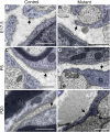
BL is fragmentary around both immature control and FAK mutant SCs in vivo. Electron micrographs of transverse sciatic nerve sections where SCs are pseudocolored to aid BL visualization. A, B, At E17.5, BL of control and mutant SCs surrounding unsorted axon bundles is thin and fragmentary, with large gaps. C, D, At P5, BL remains fragmentary around unsorted axon bundles in both control and mutant, whereas it is thicker and unbroken around thinly myelating control SCs. E, F, At P20, BL of control myelinating SCs is thick and unbroken while remaining fragmentary around unsorted axon bundles in mutants. Arrows point to BL. Scale bars, 0.5 μm. Genotypes: control, FAK fl/fl or FAKfl/+; mutant, FAKfl/fl:CNP-Cre−/+.
Figure 2.
Figure 2.
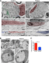
Spreading of FAK mutant SCs is impaired in vivo. A, Electron micrographs of postnatal sciatic nerves where SCs are pseudocolored to aid visualization. Transverse (a, b) and longitudinal (c–f) sections at P9 and P5, respectively, show immature SCs associated with unsorted axon bundles. Control SCs extend processes along axon bundles in both transverse (a) and longitudinal (c, e) directions. Tips of adjacent SC processes closely abut (a, e), although some regions of naked axon are observed (a, arrowhead). In contrast, mutant SCs are short and stubby and extend poorly in both transverse (b) and longitudinal (d, f) directions; extensive regions of naked axon are observed (b, d, f). Scale bars, Aa, c–f, 1 μm; Ab, 2 μm. B, Electron micrographs of transverse sections of early promyelinating SCs associated with single axons at P5. Ba, Control SCs fully envelop their associated axons, with no naked axon between the SC processes. Bb, Mutant SCs fail to envelop their associated axons, leaving extensive regions of naked axon (arrows). Bc, d, Control and mutant SCs have immature fragmentary BL (arrowhead). Regions of naked axon in mutants are associated with BL (arrow), suggesting withdrawal of SC processes. Scale bars: Ba, b, 1 μm; Bc, d, 0.25 μm. C, At P5, the percentage of sorted axons that were fully enveloped was significantly greater in control compared with mutant sciatic nerves. Values are mean ± SEM; n = 2. **p = 0.0063 (unpaired Student's t test). Genotypes: control, FAKfl/fl or FAKfl/+; mutant, FAKfl/fl:CNP-Cre−/+.
Figure 3.
Figure 3.
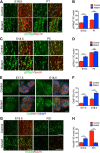
FAK mutant SCs initiate differentiation prematurely in vivo. Expression of nuclear markers of SC differentiation in transverse and longitudinal sciatic nerve sections from control and mutant mice. The sciatic nerve was delineated by immunostaining for neurofilament (NF-H) (E), or SCs were identified using the nuclear marker Sox10 (A, C, G). A, B, Longitudinal sections of sciatic nerves at E18.5 and P1. p57Kip2 is prematurely upregulated in mutant SCs at E18.5 and remains elevated compared with control at P1. Values are mean ± SEM; n = 3, 3 sections per sciatic nerve. ***p = 0.0007, E18.5 (unpaired Student's t test). ***p = 0.0005, P1 (unpaired Student's t test). C, D, Longitudinal sections of sciatic nerves at E18.5 and P5. p27Kip1 is prematurely upregulated at E18.5 in mutant SCs, but at P5 levels are not significantly different from control. Values are mean ± SEM; n = 3, 3 sections per sciatic nerve. ***p = 0.0026, E18.5 (unpaired Student's t test). ns, Not significant. E, F, Transverse sections at E17.5 and E18.5. Oct6 is prematurely upregulated at E17.5 in mutant SCs and remains elevated compared with control at E18.5. Values are mean ± SEM; n = 3, 3 sections per sciatic nerve. ***p = 0.0009, E17.5 (unpaired Student's t test). **p = 0.0079, E18.5 (unpaired Student's t test). G, H, Longitudinal and transverse sections at at E18.5 and P21, respectively. Krox20 is prematurely upregulated in mutant SCs at E18.5, but at P21 Krox20 is substantially decreased compared with control SCs. Values are mean ± SEM; n = 3 (E18.5) or 2 (P21), 3 sections per sciatic nerve. **p = 0.0038, E18.5 (unpaired Student's t test). ***p = 0.0014, P21 (unpaired Student's t test). Scale bars, 25 μm. Genotypes: control, FAKfl/fl or FAKfl/+; mutant, FAKfl/fl:CNP-Cre−/+.
Figure 4.
Figure 4.
Spreading and proliferation of SCs without functional FAK in vitro are regulated by LN concentration. Primary rat SCs expressed either EGFP (Control) or EGFP + FRNK (FRNK). A, Western blot showing that FRNK blocks endogenous FAK activity in SCs as measured by reduced phosphorylation of FAK at Y397 (autophosphorylation) and Y577 (trans-phosphorylation by Src family tyrosine kinases) and the FAK target paxillin at Y118. The potential FAK target Erk remains unaffected. Molecular masses are shown in kilodaltons. B, Phase-contrast images of control and FRNK SCs seeded on low (0.2 μg /ml) or high (5.0 μg/ml) LN for 2 and 24 h. Spreading and polarization of control SCs are complete by 24 h on both low and high LN. In contrast, FRNK SCs fail to spread or polarize on low LN by 24 h. On high LN, FRNK SCs spread but are generally unpolarized after 2 h; but by 24 h, they are spread and polarized, although less extended than control SCs. Scale bar, 100 μm. C, SCs were cultured for 24 h in the absence (−) or presence (+) of ROCK inhibitor Y27632 (5 μm). Fixed cells were incubated with rhodamine phalloidin (red) to visualize the F-actin cytoskeleton and cell shape. All SCs, except FRNK SCs on low LN, were polarized, and extended long thin processes in the presence of Y27632. FRNK SCs on low LN spread more in the presence of Y27632, although they remained round and unpolarized. Scale bar, 50 μm. D, Proliferation of SCs cultured for 24 h was measured by incubation with BrdU (10 μm) for the final 5 h. Control SCs proliferated significantly better than FRNK SCs on low LN, but Y27632 treatment normalized the proliferation of FRNK SCs. On high LN, proliferation of control and FRNK SCs was not significantly different. Values are mean ± SEM; n = 3, 2 coverslips per experiment. *p = 0.0223 (unpaired Student's t test). **p = 0.0024 (unpaired Student's t test).
Figure 5.
Figure 5.
SC differentiation is regulated by density, independently of cell-cycle status. Primary rat SCs expressed EGFP (Control), EGFP + FRNK (FRNK), or EGFP + p27Kip1 (p27Kip1). A, SCs were grown for 24 h on low (0.2 μg/ml) or high (5 μg/ml) LN and immunostained for Ki67 to identify cell-cycle status. Absence of Ki67+ SC nuclei in p27Kip1-overexpressing SCs shows that these cells are arrested in early G1. Control SCs grown on low and high LN and FRNK SCs on high LN showed similar levels of Ki67+ nuclei, whereas FRNK SCs grown on low LN had reduced levels of Ki67+ nuclei, showing that more of these cells are in early G1. Values are mean ± SEM; n = 2 separate experiments, 2 coverslips per experiment. **p = 0.0099, Control SCs low LN versus FRNK SCs low LN (unpaired Student's t test). **p = 0.0019, FRNK SCs low LN versus FRNK SCs high LN (unpaired Student's t test). ns, Not significant. B, C, dbcAMP-mediated differentiation detected by immunostaining for periaxin of control, FRNK and p27Kip1-overexpressing SCs as a function of initial cell density and LN concentration (0.2 μg/ml or 5 μg/ml; low and high LN, respectively). B, C, Immunofluorescence staining and quantitation show that, on low LN, FRNK SCs differentiated equally well when seeded at low or high density. In contrast, control SCs and p27-overexpressing SCs differentiated significantly better when grown at high density. Values are mean ± SEM; n = 3 separate experiments, 2 coverslips per experiment. **p = 0.0098 (unpaired Student's t test). ***p = 0.0007 (unpaired Student's t test). ns, Not significant. However, on high LN, each SC population showed a similar differentiation response to cell density. Values are mean ± SEM; n = 3 separate experiments, 2 coverslips per experiment. ***p = 0.0003, Control SCs 2 × 10⁴/ml compared with 40 × 104/ml (unpaired Student's t test). **p = 0.0050, FRNK SCs 2 × 10⁴/ml compared with 40 × 104/ml (unpaired Student's t test). ***p = 0.0010 p27Kip1 SCs 2 × 10⁴/ml compared with 40 × 104/ml (unpaired Student's t test). Scale bar, 100 μm.
Figure 6.
Figure 6.
FAK activity is required for SC contractilty on low LN at low cell density, and control SC contractility is reduced at high density. A–C, Primary rat SCs expressing exogenous EGFP (Control), EGFP + FRNK (FRNK), or EGFP + p27Kip1 (p27Kip1) were grown at low cell density (2 × 10⁴ cells/ml) on low or high LN. A, The F-actin cytoskeleton of SCs grown for 24 h was visualized with rhodamine phalloidin (red). Control SCs on low or high LN have prominent stress fibers arcs and lamellipodia, whereas FRNK-expressing SCs have stress fibers/arcs and lamellipodia on high but not low LN. Scale bars, 15 μm. B, C, Immunostaining for vinculin and paxillin to detect FAs. B, Control and p27Kip1-overexpressing SCs have prominent vinculin-positive FAs on low and high LN. On high LN, FRNK SCs have peripheral vinculin-positive FAs. On low LN, FRNK SCs lack vinculin-positive FAs. Scale bars, 15 μm. C, Control SCs contain prominent paxillin-positive FAs on low and high LN. FRNK SCs contain large peripheral paxillin-positive FAs on high LN, but not on low LN. Scale bars, 10 μm. D, Control SCs were seeded at 2 × 10⁴/ml (low density) or 4 × 10⁵/ml (high density) on low or high LN, then grown for 24 h before immunostaining for pMLC to detect active myosin II (red) and phalloidin staining to detect F-actin (green). SCs at low cell density have intense pMLC staining, whereas at high density immunostaining is much reduced, regardless of LN amount. Note the difference in pMLC levels between Schwann cells and the lone contaminating fibroblast in the high density culture. Phalloidin staining shows that, on low and high LN, prominent stress fibers observed at low SC density are reduced at high SC density, whereas cortical F-actin staining remains. Insets, pMLC immunostaining at low cell density. Scale bar, 100 μm.
Figure 7.
Figure 7.
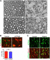
SC differentiation is not regulated by p27Kip1 or p57Kip2. A, Light microscopy of transverse sections (1 μm) of sciatic nerves stained with toluidine blue from 2-month-old mice with the following genotypes: Aa, wild-type control; Ab, FAKfl/fl:CNP-Cre−/+; Ac, p27Kip1−/−; Ad, FAKfl/fl:CNP-Cre−/+:p27Kip1−/−. Axons in p27Kip1−/− sciatic nerves are fully myelinated (Ac), and indistinguishable from control sciatic nerves (Aa). FAK mutant sciatic nerves contain large unsorted axon bundles (Ab), and ablation of p27Kip1 does not rescue this phenotype (Ad). Scale bar, 10 μm. B, Primary rat SCs expressing exogenous EGFP (control) or EGFP + p57Kip2 (p57Kip2) were seeded on high LN (5 μg/ml), differentiated for 48 h with dbcAMP (1 mm), and immunostained for periaxin (Prx) to assess differentiation. Control and p57Kip2-overexpressing Schwann cells differentiated equally well. Values are mean ± SEM; two experiments, two coverslips per experiment. ns, Not significant (unpaired Student's t test). Scale bar, 50 μm. C, P5 longitudinal sciatic nerve sections coimmunostained for p57Kip2 and Sox10 to identify SC nuclei. p57Kip2 is expressed in both mutant control SC nuclei. Scale bar, 25 μm. Genotypes: control, FAKfl/fl or FAKfl/+; mutant, FAKfl/fl:CNP-Cre−/+
Figure 8.
Figure 8.
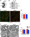
Adult SCs lacking FAK can proliferate and myelinate normally after sciatic nerve crush. Cre-mediated recombination was induced in 6-week-old control and mutant mice by tamoxifen injection. Sciatic nerves were crushed 4 months later and allowed to remyelinate. A, Genotyping of sciatic nerve genomic DNA isolated from control and mutant mice revealed highly efficient Cre-mediated recombination. After PCR amplification and digestion with HindIII, control mice had a diagnostic band of 1.9 kb, whereas after recombination in the presence of Cre-ERT2, this band was shifted to 1.1 kb. B, Western blotting of lysates from control and mutant sciatic nerves showed reduced total FAK and reduced tyrosine phosphorylation of FAK (Y397) and its target paxillin (Y118). Actin and ErbB3 were loading controls. Molecular masses are shown in kilodaltons. C, D, Control and mutant SCs proliferate similarly 7 d after crush distal to the crush site. Longitudinal sciatic nerve sections were immunostained for the nuclear markers Ki67 and Sox10. Scale bar, 100 μm. Values are mean ± SEM; n = 2; 3 sections per sciatic nerve. ns, Not significant by Student's t test. E, Electron micrographs of transverse sections of control and mutant sciatic nerves 8 weeks after crush. Comparison of control and mutant contralateral nerves shows that FAK is not required for myelin maintenance in the adult. Both control and mutant sciatic nerves were myelinated after crush, showing that FAK is not required for remyelination in the adult. Scale bar, 10 μm. F, Measurement of g-ratios in uncrushed (contralateral) and crushed (distal) control and mutant sciatic nerves 8 weeks after crush. Loss of FAK in mature sciatic nerves does not affect the g-ratios, and crushed sciatic nerves remyelinate normally. Values are mean ± SEM; n = 3 mice, 120 measurements per mouse. ns, Not significant by Student's t test. Genotypes: control mice, FAKfl/fl; mutant mice, FAKfl/fl:PLP-Cre-ERT2.

References

    1. Beggs HE, Schahin-Reed D, Zang K, Goebbels S, Nave KA, Gorski J, Jones KR, Sretavan D, Reichardt LF. FAK deficiency in cells contributing to the basal lamina results in cortical abnormalities resembling congenital muscular dystrophies. Neuron. 2003;40:501–514. doi: 10.1016/S0896-6273(03)00666-4. - DOI - PMC - PubMed
    1. Besson A, Gurian-West M, Schmidt A, Hall A, Roberts JM. p27Kip1 modulates cell migration through the regulation of RhoA activation. Genes Dev. 2004;18:862–876. doi: 10.1101/gad.1185504. - DOI - PMC - PubMed
    1. Bilodeau S, Roussel-Gervais A, Drouin J. Distinct developmental roles of cell cycle inhibitors p57Kip2 and p27Kip1 distinguish pituitary progenitor cell cycle exit from cell cycle reentry of differentiated cells. Mol Cell Biol. 2009;29:1895–1908. doi: 10.1128/MCB.01885-08. - DOI - PMC - PubMed
    1. Blanchard AD, Sinanan A, Parmantier E, Zwart R, Broos L, Meijer D, Meier C, Jessen KR, Mirsky R. Oct-6 (SCIP/Tst-1) is expressed in Schwann cell precursors, embryonic Schwann cells, and postnatal myelinating Schwann cells: comparison with Oct-1, Krox-20, and Pax-3. J Neurosci Res. 1996;46:630–640. doi: 10.1002/(SICI)1097-4547(19961201)46:5<630::AID-JNR11>3.0.CO%3B2-0. - DOI - PubMed
    1. Bryant P, Zheng Q, Pumiglia K. Focal adhesion kinase controls cellular levels of p27/Kip1 and p21/Cip1 through Skp2-dependent and -independent mechanisms. Mol Cell Biol. 2006;26:4201–4213. doi: 10.1128/MCB.01612-05. - DOI - PMC - PubMed

Publication types

MeSH terms

Substances

LinkOut - more resources