. 2014 Oct 14;111(41):14782-7.
doi: 10.1073/pnas.1417253111. Epub 2014 Oct 1.

Activation of mitochondrial protease OMA1 by Bax and Bak promotes cytochrome c release during apoptosis

Affiliations

Activation of mitochondrial protease OMA1 by Bax and Bak promotes cytochrome c release during apoptosis

Xian Jiang et al. Proc Natl Acad Sci U S A. .

Abstract

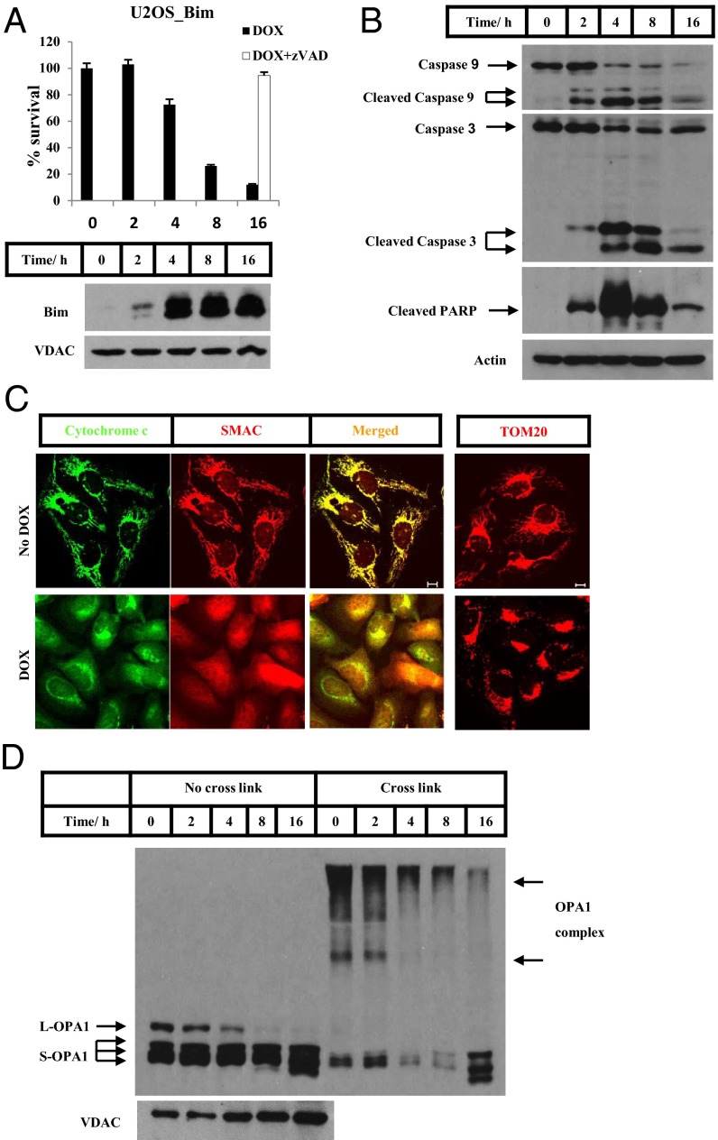
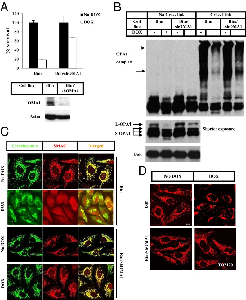
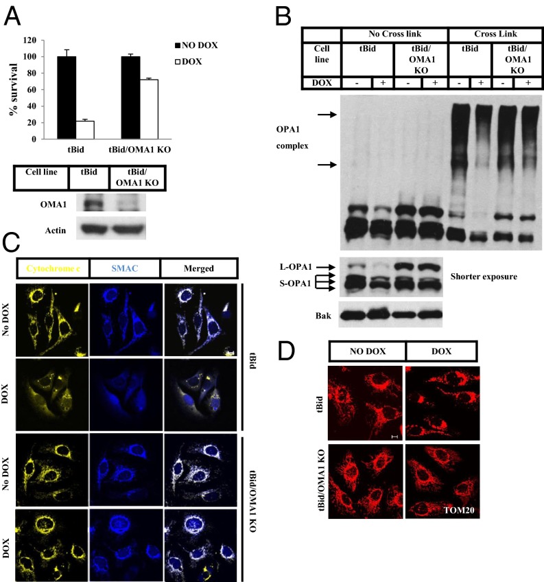
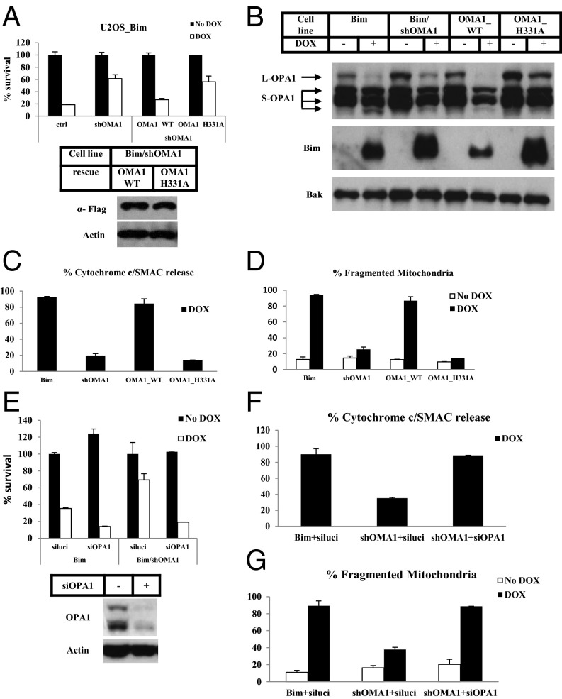
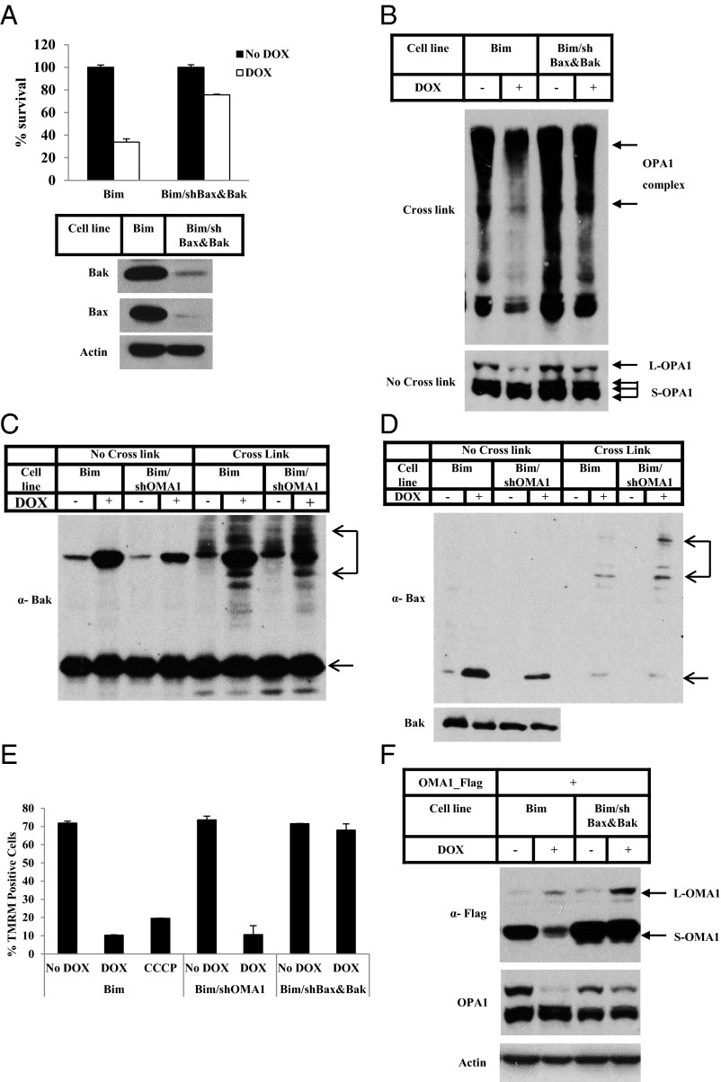
Intrinsic apoptotic stimuli initiate mammalian cells' apoptotic program by first activating the proteins that have only Bcl-2 homology domain 3 (BH3), such as Bcl-2 interacting mediator of cell death (Bim) and truncated BH3 interacting death domain agonist (tBid), which in turn trigger conformational changes in BCL2-associated X (Bax) and BCL2-antagonist/killer (Bak) proteins that enable oligomer formation on the mitochondria, causing cytochrome c and other apoptogenic proteins in the intermembrane space to leak out. Leaked cytochrome c then initiates apoptotic caspase activation through a well-defined biochemical pathway. However, how oligomerized Bax and Bak cause cytochrome c release from mitochondria remains unknown. We report here the establishment of cell lines in which Bim or tBid can be inducibly expressed to initiate apoptosis in a controlled, quantitative manner. We used these cell lines to examine apoptotic events after Bax and Bak oligomerization but before cytochrome c release. The mitochondrial metalloprotease OMA1 was activated in this system in a Bax- and Bak-dependent fashion. Activated OMA1 cleaved the dynamin-like GTPase, optical nerve atrophy 1, an event that is critical for remodeling of mitochondrial cristae. Knockdown or knockout of OMA1 in these cells attenuated cytochrome c release. Thus it is clear that oligomerized Bax and Bak trigger apoptosis by causing both the permeabilization of the mitochondrial outer membrane and activation OMA1.

Keywords: Smac; caspase; membrane potential; permeability.


Conflict of interest statement

The authors declare no conflict of interest.

Figures

Fig. 1.
Fig. 1.
Inducible expression of Bim triggers mitochondria-mediated apoptosis. (A) A U2OS cell line with a stably transfected Bim transgene (U2OS_Bim) was treated with Dox (0.1 µg/mL) in the presence or absence of z-VAD (20 µM) for the times indicated. The same concentrations were used in all experiments. Cell viability was determined by measuring ATP levels using the Cell-Titer Glo kit. Following Dox treatment for the indicated times, the P15 fractions of U2OS_Bim cells were analyzed by Western blotting. (B) U2OS_Bim cells were treated with or without Dox for the times indicated. The S15 fractions of the cells were analyzed by Western blotting. (C) U2OS_Bim cells in the presence of z-VAD were treated with or without Dox for 4 h. Immunostaining was performed using anti-cytochrome c (green) and anti-Smac (red) antibodies or anti-translocase of the outer membrane 20 (TOM20) antibody. (Scale bars: 10 µm.) (D) U2OS_Bim cells were treated with Dox for the times indicated. The P15 fractions were cross-linked with 10 mM 1,6-bis(maleimido)hexane (BMH) where indicated and were analyzed by Western blotting.
Fig. 2.
Fig. 2.
OMA1 controls Bim-induced OPA1 cleavage and disassembly of the OPA1-containing complex. (A) U2OS_Bim (Bim) cells and U2OS_Bim cells stably expressing OMA1 shRNA (Bim/shOMA1) were treated with or without Dox for 12 h. Cell viability was determined using the Cell-Titer Glo kit. Whole-cell extracts of Bim and Bim/shOMA1 cells were analyzed by Western blotting. (B) Bim and Bim/shOMA1 cells were treated with or without Dox for 8 h. The P15 fractions of the cells were cross-linked with 10 mM BMH where indicated. Western blotting was performed using the indicated antibodies. (C and D) Bim and Bim/shOMA1 cells were treated with or without Dox for 8 h. z-VAD was included during the treatment of Bim cells. Immunostaining was performed using anti-cytochrome c (green) and anti-Smac (red) antibodies (C) or anti-TOM20 antibody (D). (Scale bars: 10 µm.)
Fig. 3.
Fig. 3.
OMA1 controls tBid-induced OPA1 cleavage and disassembly of the OPA1-containing complex. (A) A U2OS cell line with a stably transfected tBid transgene (tBid) and tBid cells with stable knockout of OMA1 (tBid/OMA1 KO) were treated with or without Dox for 12 h. Cell viability was determined using the Cell-Titer Glo kit. Whole-cell extracts of tBid and tBid/OMA1 KO cells were analyzed by Western blotting. (B) tBid and tBid/OMA1 KO cells were treated with or without Dox for 8 h. The P15 fractions of the cells were cross-linked with 10 mM BMH where indicated. Western blotting was performed using the indicated antibodies. (C and D) tBid and tBid/OMA1 KO cells were treated with or without Dox for 8 h. z-VAD was included during the treatment for tBid cells. Immunostaining was performed using anti-cytochrome c (fluorescent secondary antibody with excitation at 633 nm, shown in yellow) and anti-Smac (blue) antibodies (C) or anti-TOM20 antibody (D). (Scale bar: 10 µm.)
Fig. 4.
Fig. 4.
OMA1 promotes cytochrome c release through cleavage of OPA1. (A) Bim cells (ctrl), Bim/shOMA1 cells, Bim/shOMA1 cells rescued with wild-type OMA1 (OMA1_WT), and Bim/shOMA1 cells rescued with the protease active site mutant of OMA1 (OMA1_H331A) were treated with or without Dox for 16 h. Cell viability was determined using the Cell-Titer Glo kit. Whole-cell extracts of the indicated cell lines were analyzed by Western blotting. (B) The indicated cell lines were treated with or without Dox for 16 h. The P15 fractions of the cells were analyzed by Western blotting. (C and D) The indicated cell lines were treated with or without Dox for 16 h. z-VAD was included during the treatment for Bim and OMA1_WT cells. Percentages of cells with cytochrome c and Smac release into cytosol (C) or percentages of cells with fragmented mitochondria (D) as examined by immunostaining were calculated. Mitochondria were visualized with immunostaining of TOM20. (E) Bim and Bim/shOMA1 cells were transfected with siRNA against luciferase (luci) or OPA1. Forty-eight hours later, cells were treated with or without Dox for 12 h. Cell viability was determined using the Cell-Titer Glo kit. Whole-cell extracts of Bim cells before and after 48 h of OPA1 siRNA transfection were analyzed by Western blotting. (F and G) Bim and Bim/shOMA1 cells were transfected with the indicated siRNAs and were treated with or without Dox for 8 h. z-VAD was included during the treatment for Bim cells and Bim/shOMA1 cells with OPA1 siRNA transfection. Percentages of cells with cytochrome c and Smac release into cytosol (F) and percentages of cells with fragmented mitochondria (G) as determined by immunostaining were calculated.
Fig. 5.
Fig. 5.
Bax and Bak function upstream of OMA1 activation. (A) Bim cells and Bim cells stably expressing both Bax and Bak shRNAs (Bim/shBax&Bak) were treated with or without Dox for 12 h. Cell viability was determined using the Cell-Titer Glo kit. Whole-cell extracts of Bim and Bim/shBax&Bak cells were analyzed by Western blotting. (BD) Bim and Bim/shBax&Bak cells (B) or Bim and Bim/shOMA1 cells (C and D) were treated with or without Dox for 8 h. The P15 fractions were cross-linked with 10 mM BMH where indicated. Western blotting was performed using the indicated antibodies. (E) The indicated cell lines were treated with or without Dox for 8 h or were treated with 10 µM CCCP for 90 min. TMRM staining followed by flow cytometry analysis was performed. TMRM+ cells were quantified. (F) Bim and Bim/shBax&Bak cells were transfected with Flag-tagged OMA1 for 48 h. Cells then were treated with Dox for 8 h or were left untreated. Whole-cell extracts were analyzed by Western blotting. L-OMA1, full-length precursor OMA1; S-OMA1, shorter version of OMA1.

References

    1. Liu X, Kim CN, Yang J, Jemmerson R, Wang X. Induction of apoptotic program in cell-free extracts: Requirement for dATP and cytochrome c. Cell. 1996;86(1):147–157. - PubMed
    1. Li P, et al. Cytochrome c and dATP-dependent formation of Apaf-1/caspase-9 complex initiates an apoptotic protease cascade. Cell. 1997;91(4):479–489. - PubMed
    1. Du C, Fang M, Li Y, Li L, Wang X. Smac, a mitochondrial protein that promotes cytochrome c-dependent caspase activation by eliminating IAP inhibition. Cell. 2000;102(1):33–42. - PubMed
    1. Verhagen AM, et al. Identification of DIABLO, a mammalian protein that promotes apoptosis by binding to and antagonizing IAP proteins. Cell. 2000;102(1):43–53. - PubMed
    1. Kluck RM, Bossy-Wetzel E, Green DR, Newmeyer DD. The release of cytochrome c from mitochondria: A primary site for Bcl-2 regulation of apoptosis. Science. 1997;275(5303):1132–1136. - PubMed

Publication types

MeSH terms

Substances