Review
. 2015 Jan;14(1):50-60.
doi: 10.1093/bfgp/elu038. Epub 2014 Sep 30.

Functional roles of nucleosome stability and dynamics

Review

Functional roles of nucleosome stability and dynamics

Răzvan V Chereji et al. Brief Funct Genomics. 2015 Jan.

Abstract

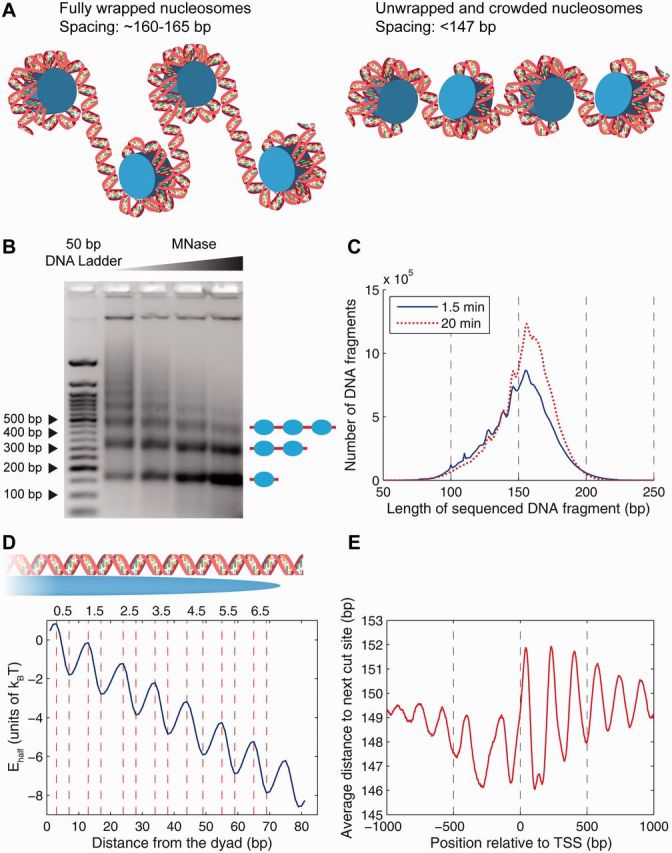
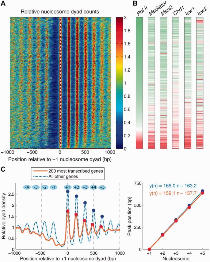
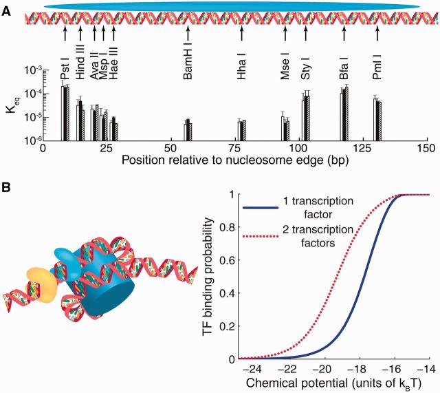
Nucleosome is a histone-DNA complex known as the fundamental repeating unit of chromatin. Up to 90% of eukaryotic DNA is wrapped around consecutive octamers made of the core histones H2A, H2B, H3 and H4. Nucleosome positioning affects numerous cellular processes that require robust and timely access to genomic DNA, which is packaged into the tight confines of the cell nucleus. In living cells, nucleosome positions are determined by intrinsic histone-DNA sequence preferences, competition between histones and other DNA-binding proteins for genomic sequence, and ATP-dependent chromatin remodelers. We discuss the major energetic contributions to nucleosome formation and remodeling, focusing especially on partial DNA unwrapping off the histone octamer surface. DNA unwrapping enables efficient access to nucleosome-buried binding sites and mediates rapid nucleosome removal through concerted action of two or more DNA-binding factors. High-resolution, genome-scale maps of distances between neighboring nucleosomes have shown that DNA unwrapping and nucleosome crowding (mutual invasion of nucleosome territories) are much more common than previously thought. Ultimately, constraints imposed by nucleosome energetics on the rates of ATP-dependent and spontaneous chromatin remodeling determine nucleosome occupancy genome-wide, and shape pathways of cellular response to environmental stresses.

Keywords: DNA; chromatin; gene regulation; nucleosome; stress response; transcription factor.


Figures

Figure 1:
Figure 1:
Energetics of DNA unwrapping and nucleosome spacing in the yeast genome. (A) Left panel: fully wrapped nucleosomes with ∼160–165 bp spacing between neighboring dyads. Right panel: partially unwrapped and crowded nucleosomes with <147 bp spacing between neighboring dyads. (B) Nucleosome ladder on an agarose gel. Wild-type yeast was grown in synthetic complete medium. DNA purified from nuclei digested with increasing amounts of MNase was analyzed in a 2% agarose gel stained with ethidium bromide. Leftmost lane: 50 bp DNA ladder (New England Biolabs). Image courtesy of Drs Josefina Ocampo and David J. Clark (NICHD, NIH). (C) Histogram of DNA fragment lengths from the high-resolution chemical map, 1.5 min (blue, solid) and 20 min (red, dotted) after the addition of hydrogen peroxide [8]. (D) Histone–DNA unwrapping and higher-order structure energy profile. The total energy of a partially wrapped nucleosome with x bp of DNA to the left of the dyad and y bp of DNA to the right of the dyad is given by Ehalf(x)+Ehalf(y). The minima and maxima of the energy landscape are based on crystal structures of nucleosome core particles [9, 10]. Histone–DNA contact patches, corresponding to the regions where the minor groove of the DNA double helix faces inward, are labeled 0.5, 1.5, 2.5, 3.5, 4.5, 5.5 and 6.5. (E) Average distance between hydroxyl cut sites marking neighboring dyads in the vicinity of the TSS. The genome-wide average distance is 150.9 bp. (A colour version of this figure is available online at: http://bfg.oxfordjournals.org)
Figure 2:
Figure 2:
Chromatin organization, gene transcription and remodeler activity in S. cerevisiae. (A) Heatmap of nucleosome dyad counts in S. cerevisiae in the vicinity of coding regions; genes are sorted by Pol II occupancy averaged in the [TSS,TTS] range (TTS: transcription termination site; nucleosome and Pol II data: wild-type cells in glucose [47]). (B) DNA occupancy levels for Pol II, Mediator, TF Msn2 [47] and chromatin remodelers Chd1, Isw1, Isw2 [48]. Genes are sorted as in (A). Pol II and Mediator: wild-type yeast in glucose; Msn2: wild-type yeast 20 min after a glucose-to-glycerol switch; Chd1, Isw1, Isw2: wild-type yeast grown in yeast extract peptone dextrose (YPD) medium [48]. For each factor, the occupancy was averaged in the [TSS-300, TSS] range (Mediator, Msn2), or in the [TSS, TTS] range (Pol II, Chd1, Isw1, Isw2). Distributions of average occupancies over all yeast genes were converted into z-scores, and the color scheme in each vertical bar was set so that genes in the bottom 5 percentile (negative z-scores) are green, genes in the top 5 percentile (positive z-scores) are red and genes with zero z-scores are white. (C) Left panel: average nucleosome dyad density in the 200 most transcribed genes (orange, solid) and in the rest of the yeast genes (blue, dotted). Dots mark peaks of nucleosome dyad density in the coding region. Right panel: linear fit to the positions of the nucleosome density peaks shown in the left panel. The slope of the fit is an estimate of the inter-nucleosome spacing based on single-nucleosome data. (A colour version of this figure is available online at: http://bfg.oxfordjournals.org)
Figure 3:
Figure 3:
DNA accessibility and TF binding to nucleosomal DNA. (A) Restriction enzyme sites inserted into the 601 nucleosome sequence at locations indicated by black arrows [56]. Each group of three bars represents independent measurements in which the 601 sequence was flanked by different DNA sequences. The height of each bar is the equilibrium constant Keq for site exposure averaged over multiple experiments; error bars show standard deviations. (B) Nucleosome-induced cooperativity between two DNA-binding factors. Left panel: two TFs bound simultaneously to nucleosomal DNA; the nucleosome is partially unwrapped. Right panel: TF binding probability at a site located at bp 31–40 from the edge of the fully wrapped nucleosome, in the absence (blue, solid) or presence (red, dotted) of a second site for the same TF located on the same side of the dyad, at bp 11–20. The free energy of a fully wrapped nucleosome is ln(109) kBT; histone chemical potential is ln(106) kBT; TF binding energy is ln(1010) kBT for its cognate sites and ln(106) kBT for all other sites [57]. The model of nucleosome unwrapping is as described in [42]. (A colour version of this figure is available online at: http://bfg.oxfordjournals.org)

References

    1. Grosberg AY, Khokhlov AR. Statistical Physics of Macromolecules. Woodbury, NY: AIP Press; 1994.
    1. Frank-Kamenetskii MD. Biophysics of the DNA molecule. Phys Rep. 1997;288:13–60.
    1. van Holde KE. Chromatin. New York: Springer; 1989.
    1. Arents G, Moudrianakis EN. Topography of the histone octamer surface: repeating structural motifs utilized in the docking of nucleosomal DNA. Proc Natl Acad Sci USA. 1993;90:10489–93. - PMC - PubMed
    1. McBryant SJ, Adams VH, Hansen JC. Chromatin architectural proteins. Chromosome Res. 2006;14:39–51. - PubMed

Publication types