

Oncocytoma-like renal tumor with transformation toward high-grade oncocytic carcinoma: a unique case with morphologic, immunohistochemical, and genomic characterization
- PMID: 25275525
- PMCID: PMC4616290
- DOI: 10.1097/MD.0000000000000081
Oncocytoma-like renal tumor with transformation toward high-grade oncocytic carcinoma: a unique case with morphologic, immunohistochemical, and genomic characterization
Abstract
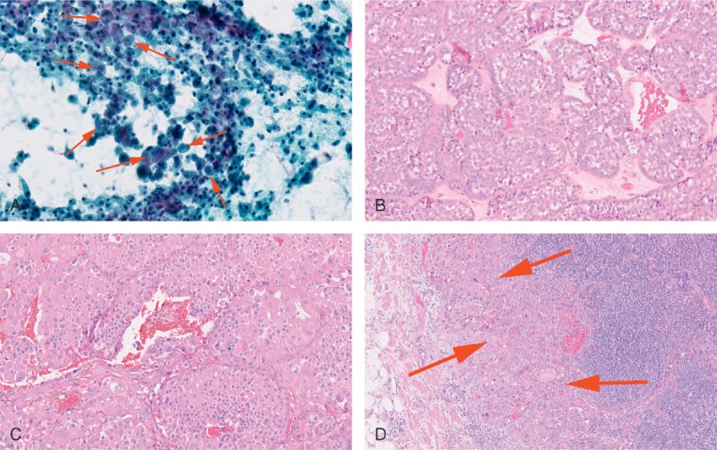
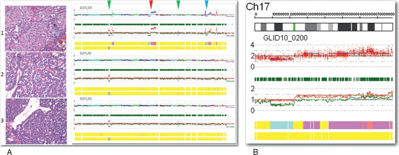
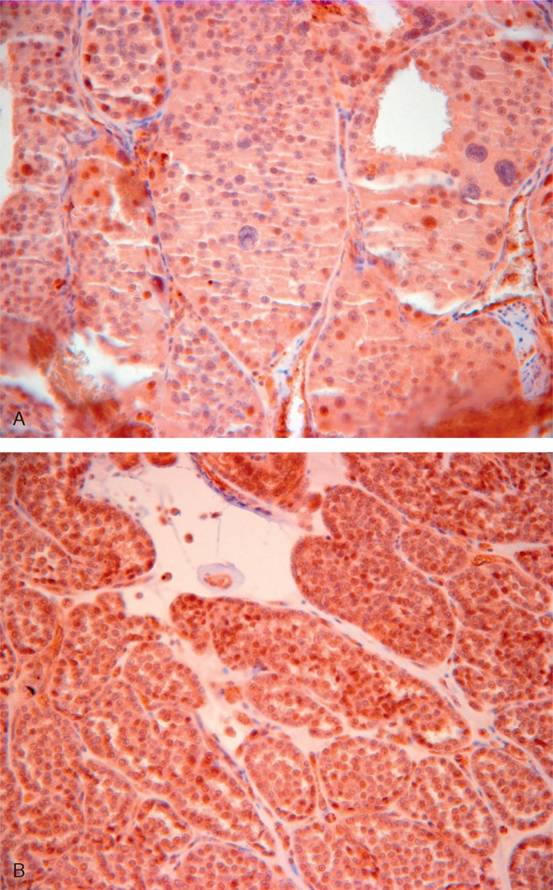
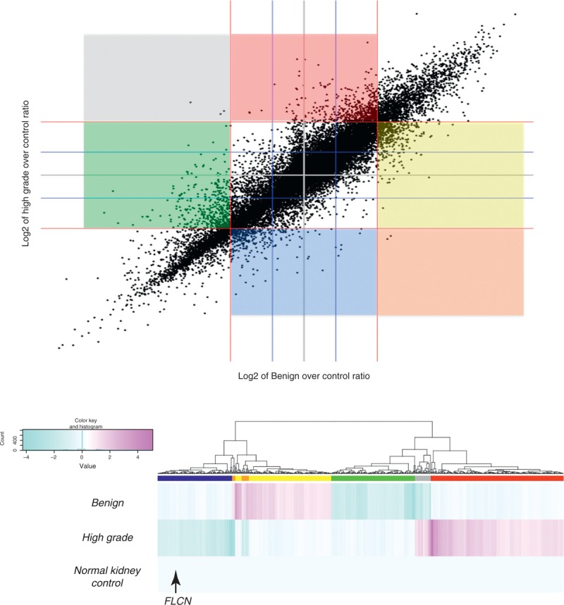
Renal oncocytoma is a benign tumor with characteristic histologic findings. We describe an oncocytoma-like renal tumor with progression to high-grade oncocytic carcinoma and metastasis. A 74-year-old man with no family history of cancer presented with hematuria. Computed tomography showed an 11 cm heterogeneous multilobulated mass in the right kidney lower pole, enlarged aortocaval lymph nodes, and multiple lung nodules. In the nephrectomy specimen, approximately one third of the renal tumor histologically showed regions classic for benign oncocytoma transitioning to regions of high-grade carcinoma without sharp demarcation. With extensive genomic investigation using single nucleotide polymorphism-based array virtual karyotyping, multiregion sequencing, and expression array analysis, we were able to show a common lineage between the benign oncocytoma and high-grade oncocytic carcinoma regions in the tumor. We were also able to show karyotypic differences underlying this progression. The benign oncocytoma showed no chromosomal aberrations, whereas the high-grade oncocytic carcinoma showed loss of the 17p region housing FLCN (folliculin [Birt-Hogg-Dubé protein]), loss of 8p, and gain of 8q. Gene expression patterns supported dysregulation and activation of phosphoinositide 3-kinase (PI3K)/v-akt murine thymoma viral oncogene homolog (Akt), mitogen-activated protein kinase (MAPK)/extracellular-signal-regulated kinase (ERK), and mechanistic target of rapamycin (serine/threonine kinase) (mTOR) pathways in the high-grade oncocytic carcinoma regions. This was partly attributable to FLCN underexpression but further accentuated by overexpression of numerous genes on 8q. In the high-grade oncocytic carcinoma region, vascular endothelial growth factor A along with metalloproteinases matrix metallopeptidase 9 and matrix metallopeptidase 12 were overexpressed, facilitating angiogenesis and invasiveness. Genetic molecular testing provided evidence for the development of an aggressive oncocytic carcinoma from an oncocytoma, leading to aggressive targeted treatment but eventual death 39 months after the diagnosis.
Conflict of interest statement
The authors have no funding and conflicts of interest to disclose.
AG, RPB, and ZG are employees of Caris Life Sciences (Phoenix, AZ and Dallas, TX); BLL is an employee of Transgenomic, Inc. (Omaha, NE). The other authors have no conflicts of interest to disclose.
Figures
References
-
- Monzon FA, Hagenkord JM, Lyons-Weiler MA, et al. Whole genome SNP arrays as a potential diagnostic tool for the detection of characteristic chromosomal aberrations in renal epithelial tumors. Mod Pathol. 2008;21:599–608. - PubMed
-
- Hagenkord JM, Gatalica Z, Jonasch E, et al. Clinical genomics of renal epithelial tumors. Cancer Genet. 2011;204:285–297. - PubMed
-
- Presti JC, Jr, Wilhelm M, Reuter V, et al. Allelic loss on chromosomes 8 and 9 correlates with clinical outcome in locally advanced clear cell carcinoma of the kidney. J Urol. 2002;167:1464–1468. - PubMed
-
- Junker K, Romics I, Szendroi A, et al. Genetic profile of bone metastases in renal cell carcinoma. Eur Urol. 2004;45:320–324. - PubMed
-
- Klatte T, Kroeger N, Rampersaud EN, et al. Gain of chromosome 8q is associated with metastases and poor survival of patients with clear cell renal cell carcinoma. Cancer. 2012;118:5777–5782. - PubMed
Publication types
MeSH terms
Substances
Supplementary concepts
LinkOut - more resources
Full Text Sources
Other Literature Sources
Medical
Miscellaneous
